Method and device for segmenting a medical examination object with quantitative magnetic resonance imaging

a medical examination object and quantitative magnetic resonance imaging technology, applied in the field of quantitative magnetic resonance imaging methods and devices, can solve the problems of prone to errors, further image differences, and qualitative contrast, and achieve the effect of avoiding the drawbacks of qualitative methods

Active Publication Date: 2016-06-02
SIEMENS HEALTHCARE GMBH
View PDF3 Cites 14 Cited by
  • Summary
  • Abstract
  • Description
  • Claims
  • Application Information

AI Technical Summary

Benefits of technology

[0031]The user of the inventive method and the associated device is therefore advantageously capable of carrying out segmenting ...

Problems solved by technology

Current segmenting algorithms for structures having complex geometries, such as e.g. joints, are often computing-intensive and in particular in conventional MR images, which generate only one qualitative contrast, prone to error.
Errors can occur during segmenting if the scan protocol is changed too much.
Further image differences can occur due to the patient positio...

Method used


Image

  • Method and device for segmenting a medical examination object with quantitative magnetic resonance imaging
  • Method and device for segmenting a medical examination object with quantitative magnetic resonance imaging
  • Method and device for segmenting a medical examination object with quantitative magnetic resonance imaging

Examples


Embodiment Construction

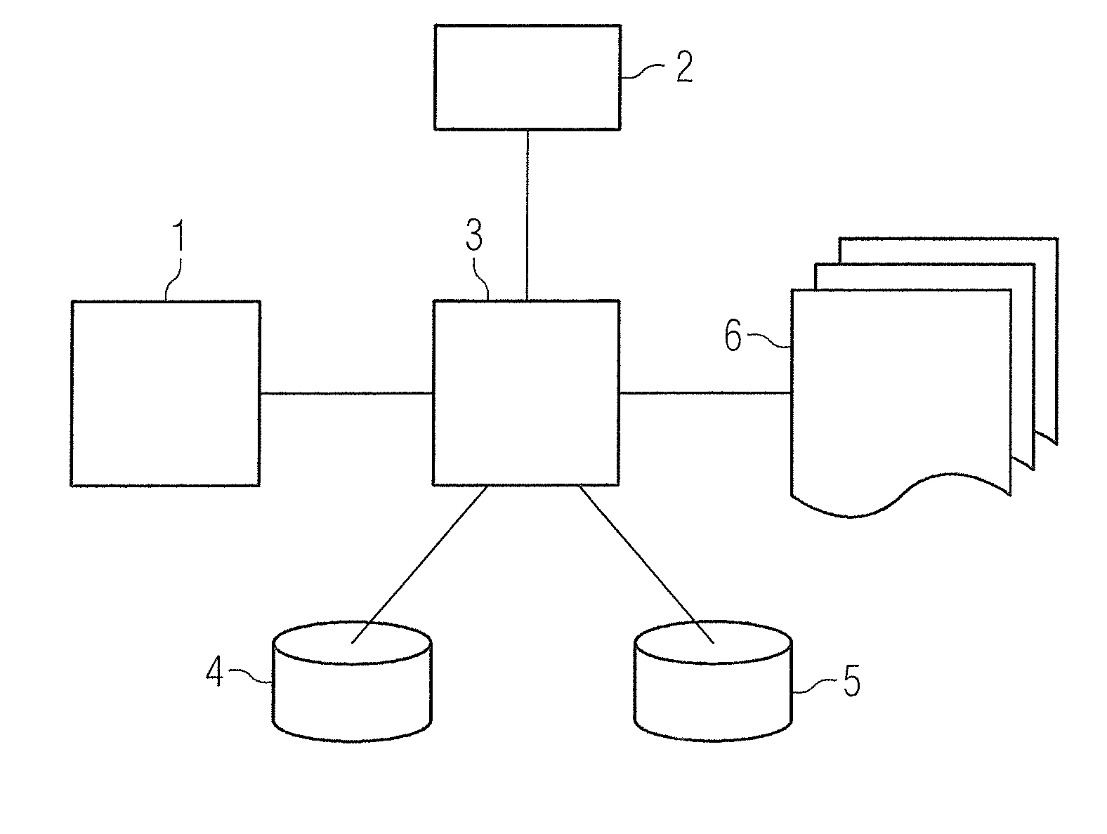
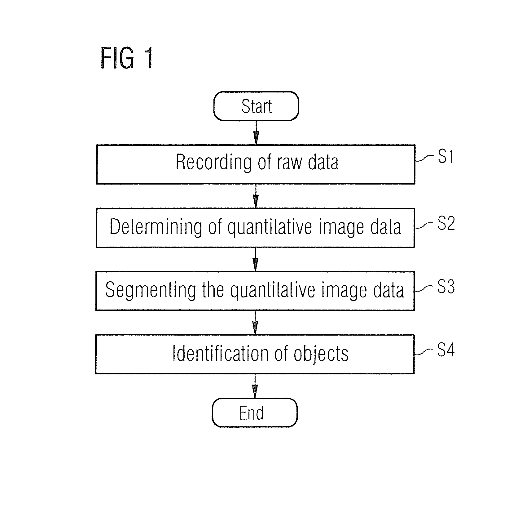
[0035]FIG. 1 shows method steps for segmenting image data of an examination object. In the first step S1, the raw data of the examination object are acquired. The raw data include digitized MR signals that are produced during the scan sequence of the examination object. The signals are stored in a matrix (k-space) in a memory. The data stored in this way can be converted by a Fourier transformation for further processing.

[0036]The above-described raw data can include a large number of different items of information about the tissue. After the above-described transformation these items of information are available in the form of quantitative tissue parameters which are spatially and / or temporally resolved and include specific regions of the examination object or even the entire examination object.

[0037]These tissue parameters are combined in step S2 to form quantitative image data, it being possible for this to include for example T1, T2 or the spin density. T1 denotes the relaxation...


PUM


Abstract

In a method and magnetic resonance apparatus for segmenting image data of an examination object, raw data of the examination object are achieved with the operation of a magnetic resonance scanner. Quantitative image data of the examination object are then calculated in a processor from the raw data. At least one physical variable of the examination object is quantitatively ascertained pixelwise and is displayed. The quantitative image data are segmented for identification of predetermined objects in the quantitative image data, and displayed in a display unit.

Description

BACKGROUND OF THE INVENTION[0001]1. Field of the Invention[0002]The invention concerns a method and a device for segmenting a medical examination object by the application of quantitative magnetic resonance (MR) imaging methods for medical issues and segmenting processes.[0003]2. Description of the Prior Art[0004]The significance of segmenting processes, i.e. the depiction of specific sections of examination objects, has increased greatly in the past years in MR imaging in general and in orthopedic MR imaging in particular. One application example is the segmenting of cartilage tissue and the subsequent automatic evaluation of thickness, volume, etc. to detect potential cartilage damage as early as possible and to objectively quantify a potential successful treatment or course over time of diseases (e.g. arthroses). Other areas are e.g. segmenting of tumors or vessel deposits. Segmenting methods are also used to depict bones or fluids or in MR PET attenuation correction. Current seg...

Claims


Application Information

Patent Timeline
IPC IPC(8): G06T7/00A61B5/055A61B5/00G06K9/00
CPCG06T7/0097G06T7/0012G06K9/00885A61B5/055G06K2009/00939G06T2207/10088G06T2207/10024G06T2207/30024A61B5/743A61B5/0037A61B2576/00G01R33/5608G06T7/0014G06T2207/20036G06T2207/30008G06T7/11G06T7/62G16H30/40
Inventor BACHSCHMIDT, THERESAGRODZKI, DAVIDMEYER, HEIKONITTKA, MATHIASRAITHEL, ESTHER
Owner SIEMENS HEALTHCARE GMBH
Why Eureka
Try Eureka