Image quality of a magnetic resonance image dataset

Active Publication Date: 2016-07-28
SIEMENS HEALTHCARE GMBH
View PDF2 Cites 7 Cited by
  • Summary
  • Abstract
  • Description
  • Claims
  • Application Information

AI Technical Summary

Benefits of technology

[0010]In one embodiment, an iterative image quality improvement method for magnetic resonance imaging is provided, which is applied in the k-space and uses correction image datasets of another modality, (e.g., x-ray image datasets), in order to optimize the image quality of magnetic resonance image datasets. Thus, the information that was obtained by another imaging modality is ultimately used to be able to compensate at least partly for limitations in the magnetic resonance imaging. Use is made of the fact that the correction image datasets contain information about the materials in the recording area, so that these are able to be classified with respect to the magnetic resonance imaging, thus are able to be described in their magnetic characteristics, which in turn makes it possible to estimate that result the magnetic resonance imaging may have produced under idealized conditions. This result is created in the form of the virtual magnetic resonance comparison dataset, which ultimately represents a kind of orientation point that may show where and/or how corrections are to be made to the magnetic resonance image dataset in order to improve the image quality with respect to limitations in the magnetic resonance imaging that are not available with the other modality. Thus, the comparison of the magnetic resonance image dataset and the magnetic resonance comparison dataset, which may also be carried out at least partly in the k-space, as will be discussed in greater detail below, makes it possible to determine measure parameters, (e.g., filter parameters, interpolation parameters, distortion removal parameters, and the like), in order to carry out concrete image quality improvement measures. These are applied in the k-space, since inadequacies or limitations in the magnetic resonance imaging origina

Problems solved by technology

This localization may, however, be lost in the local space, so tha

Method used


Image

  • Image quality of a magnetic resonance image dataset
  • Image quality of a magnetic resonance image dataset
  • Image quality of a magnetic resonance image dataset

Examples


Embodiment Construction

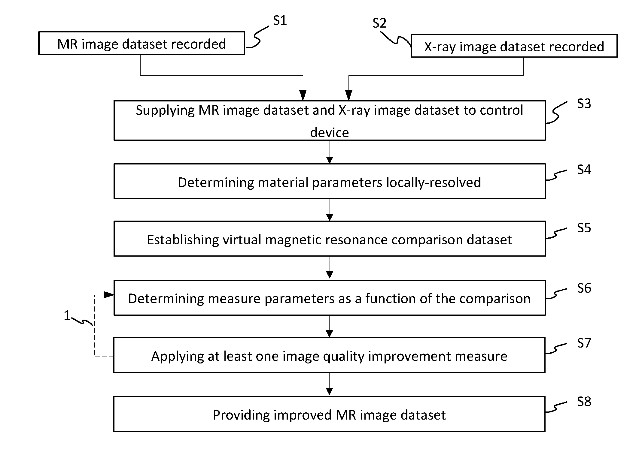
[0039]FIG. 1 depicts a flowchart of an exemplary embodiment of the method, as may be carried out on a combination imaging device that combines a magnetic resonance device and an x-ray device. The combination imaging device allows simultaneous recording of magnetic resonance image datasets and x-ray image datasets, wherein, in the method presented in more detail here, a three-dimensional x-ray image dataset is used for improving the image quality of a three-dimensional magnetic resonance image dataset, for example, as part of angiographic examinations.

[0040]Thus, in corresponding acts S1 and S2, the magnetic resonance image dataset and the x-ray image dataset are recorded with the combination-imaging device and are then supplied to a control device of the combination-imaging device for joint processing, act S3. There, the x-ray image dataset, which in this exemplary embodiment represents the correction image dataset, is now initially used, in act S4, in order, at least for the record...


PUM


Abstract

A method for improving the image quality of a three-dimensional magnetic resonance image dataset recorded with a magnetic resonance device, wherein, from at least one correction image dataset recorded with a modality other than magnetic resonance imaging, registered with the magnetic resonance image dataset, showing at least partly the same recording region as the magnetic resonance image dataset, especially an x-ray image dataset, relevant material parameters are derived locally-resolved for the magnetic resonance imaging, which are used for establishing a virtual magnetic resonance comparison dataset in a simulation wherein, as a function of a comparison between the magnetic resonance image dataset and the magnetic resonance comparison dataset, at least one measure parameter describing an image quality improvement measure to be applied in the k-space is determined and the image quality improvement measure is carried out with the measure parameter relating to the magnetic resonance image dataset.

Description

[0001]This application claims the benefit of DE 10 2015 201 057.0, filed on Jan. 22, 2015, which is hereby incorporated by reference in its entirety.TECHNICAL FIELD[0002]The embodiments relate to a method for improving the image quality of a three-dimensional magnetic resonance image dataset recorded with a magnetic resonance device, a computing device, and a computer program.BACKGROUND[0003]Magnetic resonance imaging has become established as a modality for the examination of patients in medical devices, since it provides a number of advantages, such as the outstanding soft tissue contrast of magnetic resonance imaging. There are limitations in relation to magnetic resonance imaging, however, which to some extent are inherent, but are also caused to some extent by the specific circumstances for recording a magnetic resonance image dataset. These limitations are at the expense of the image quality of the magnetic resonance image dataset. For example, recordings with high local resol...

Claims


Application Information

Patent Timeline
IPC IPC(8): G06T5/00G06T15/00G06K9/52G06T7/60G06T7/00G06K9/62
CPCG06T5/00G06T7/0012G06K9/6201G06K9/52G06T7/60G06T2207/20024G06T15/005G06T2200/04G06T2207/10088G06T2207/30168G06T7/0024G06T19/00G01R33/4812G01R33/5608G01R33/565
Inventor ERTEL, DIRKKYRIAKOU, YIANNIS
Owner SIEMENS HEALTHCARE GMBH
Why Eureka
Try Eureka