Method and System for Advanced Transcatheter Aortic Valve Implantation Planning

a transcatheter aortic valve and advanced technology, applied in the field of advanced transcatheter aortic valve implantation planning, can solve the problem of not being able to assess only, and achieve the effect of reducing the number of patients

Active Publication Date: 2016-10-20
SIEMENS HEALTHCARE GMBH
View PDF7 Cites 32 Cited by
  • Summary
  • Abstract
  • Description
  • Claims
  • Application Information

AI Technical Summary

Benefits of technology

[0003]The present invention provides a method and system for transcatheter aortic valve implantation (TAVI) planning. Embodiments of the present invention provide an automated framework to extract geometric models of the aortic valve, including calcium, from operative medical image data, that can be printed using a 3D single or multi-material printer to create a physical 3D model of a patient's aortic valve. The final printed 3D model can be created using two different materials representing the two main tissues within valves of stenotic patients of aortic tissue (softer tissue) and calcium (stiffer tissue). Embodiments of the present invention allow for a seamless and fast workflow to go from medical images (e.g., CT images) to 3D printed models within minimal or no user interaction. Based on the printed 3D model of the patient's aortic valve, hands-on preoperative planning for TAVI procedures can be performed, including trying different types of implant devices and different sizes for each type of implant device.

Problems solved by technology

In addition, due to calcium within the aortic valve, it may not be enough to assess solely the geometric properties of the patient's aortic valve in order to select the right implant device.

Method used


Image

  • Method and System for Advanced Transcatheter Aortic Valve Implantation Planning
  • Method and System for Advanced Transcatheter Aortic Valve Implantation Planning
  • Method and System for Advanced Transcatheter Aortic Valve Implantation Planning

Examples


Embodiment Construction

[0016]The present invention relates to transcatheter aortic valve implantation (TAVI) planning. Embodiments of the present invention are described herein to give a visual understanding of the TAVI planning method. A digital image is often composed of digital representations of one or more objects (or shapes). The digital representation of an object is often described herein in terms of identifying and manipulating the objects. Such manipulations are virtual manipulations accomplished in the memory or other circuitry / hardware of a computer system. Accordingly, is to be understood that embodiments of the present invention may be performed within a computer system using data stored within the computer system.

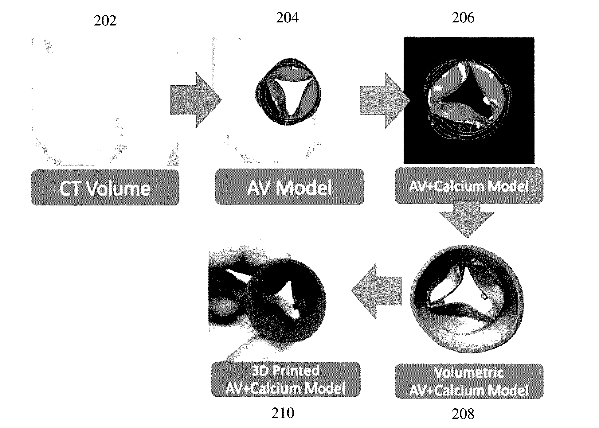
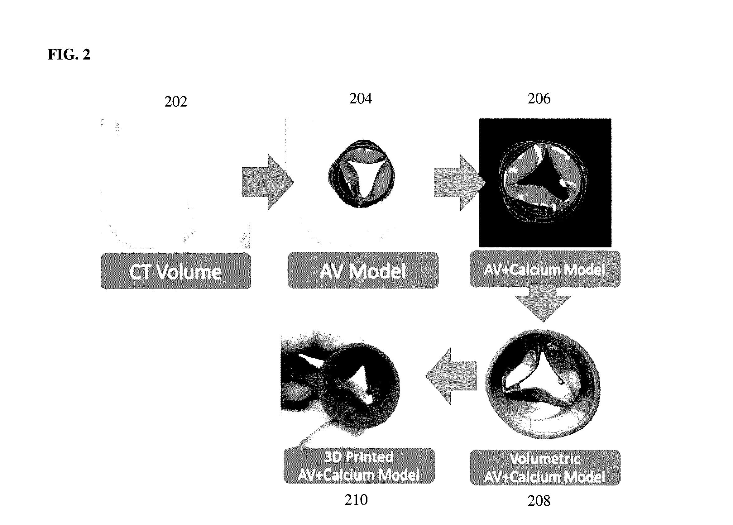
[0017]Embodiments of the present invention provide an automated framework to extract geometric models of the aortic valve, including calcium, from operative medical image data, that can be printed using a 3D single or multi-material printer to create a physical 3D model of a patien...


PUM

PropertyMeasurementUnit
surface modelaaaaaaaaaa
stiffnessaaaaaaaaaa
stiffness propertyaaaaaaaaaa

Abstract

A method and system for transcatheter aortic valve implantation (TAVI) planning is disclosed. An anatomical surface model of the aortic valve is estimated from medical image data of a patient. Calcified lesions within the aortic valve are segmented in the medical image data. A combined volumetric model of the aortic valve and calcified lesions is generated. A 3D printed model of the heart valve and calcified lesions is created using a 3D printer. Different implant device types and sizes can be placed into the 3D printed model of the aortic valve and calcified lesions to select an implant device type and size for the patient for a TAVI procedure. The method can be similarly applied to other heart valves for any type of heart valve intervention planning.

Description

BACKGROUND OF THE INVENTION[0001]The present invention relates to transcatheter aortic valve implantation (TAVI) planning, and more particularly, to generating a personalized 3D-printed anatomical model of the aortic valve for planning a TAVI procedure.[0002]Transcatheter aortic valve implantation (TAVI) is becoming the standard choice of care for non-operable and high-risk patients suffering from severe aortic valve stenosis. TAVI is a minimally invasive cardiac intervention in which an aortic valve implant is delivered into a patient through the patient's vessels via a catheter. As there is no direct view or access to the affected anatomy in a TAVI procedure, accurate pre-operative planning is crucial for a successful outcome. Computed tomography (CT) has been established as the gold standard imaging modality for pre-operative planning for TAVI procedures. Standard clinical measurements, such as annular diameters and hinge-annulus plane distance can be estimated based on geometric...

Claims


Application Information

Patent Timeline
Patent Type & Authority Applications(United States)
IPC IPC(8): B29C67/00G05B19/4099A61F2/24
CPCB29C67/0088A61F2/2496B33Y50/02G05B2219/35134G05B2219/49007G05B19/4099A61F2/2472A61F2240/00A61F2240/008B33Y80/00A61B5/1076A61B2576/023A61F2/2412A61F2/2415A61F2240/002B29C64/386B33Y50/00G06T7/0012G06T7/11G06T7/136G06T7/75G06T19/00G06T2207/10081G06T2207/20081G06T2207/30048G09B23/285G16H30/40
Inventor GRBIC, SASAIONASEC, RAZVANMANSI, TOMMASOVOIGT, INGMARNEUMANN, DOMINIKKREBS, JULIANSCHWEMMER, CHRISSCHOEBINGER, MAXHOULE, HELENE C.COMANICIU, DORINMANCINA, JOEL
Owner SIEMENS HEALTHCARE GMBH
Why Eureka
Try Eureka