Method for analyzing markers on the surface of vesicles
a technology of vesicles and markers, applied in the field of cell biology, can solve the problem of not divulging a method for analyzing the level of markers of subpopulations of extracellular vesicles
- Summary
- Abstract
- Description
- Claims
- Application Information
AI Technical Summary
Benefits of technology
Problems solved by technology
Method used
Image
Examples
case 2
er is Present Only on a Vesicle Subpopulation
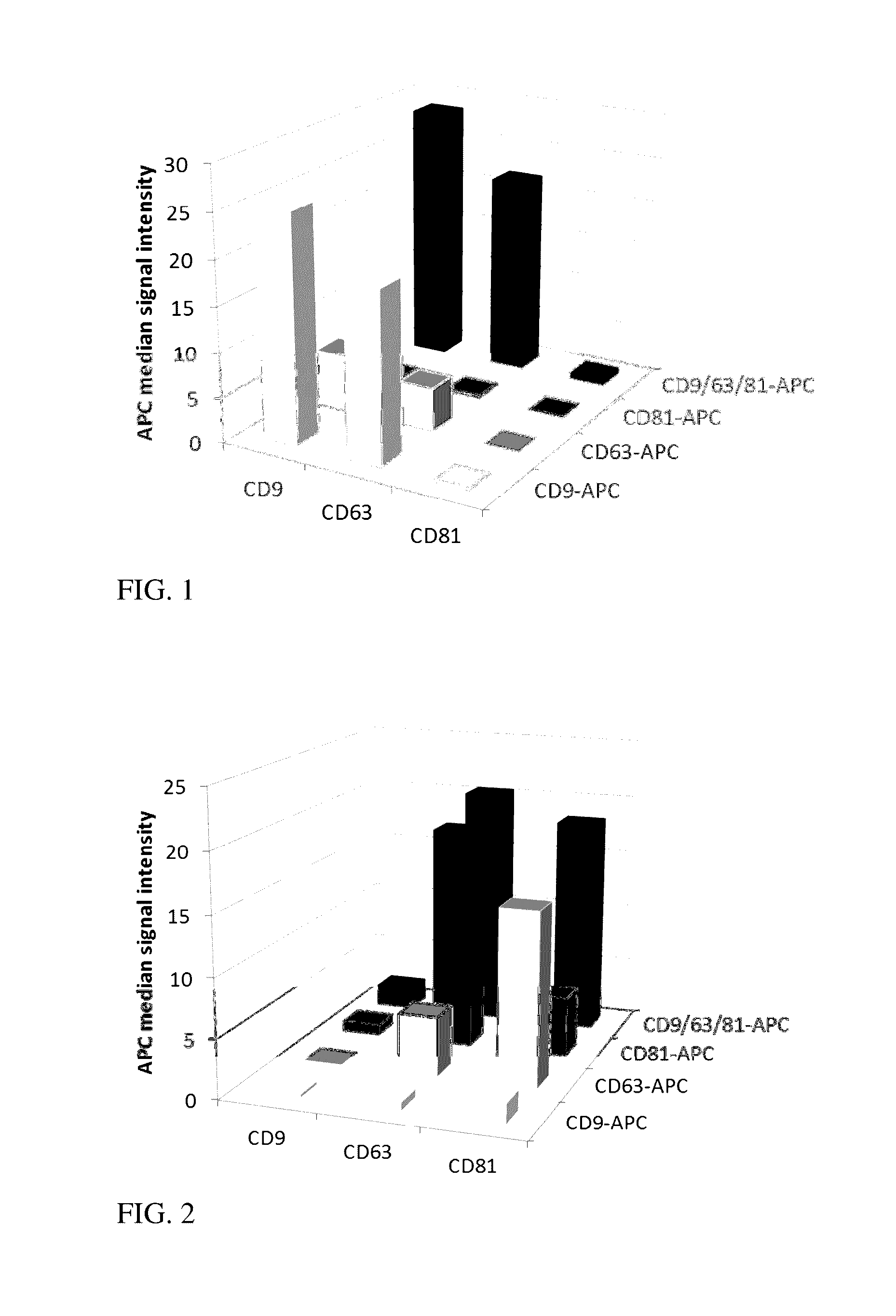
[0069]Antibody B or a fragment thereof recognizes a marker B present only on a subpopulation of the vesicles, but is abundant on the positive vesicles. The combination of A beads with B antibody detection will give a bright signal, yet lower as the AA combination as only a subset of bound vesicles is detected. The other way round, B beads will bind only a fraction of all vesicles (most of the vesicles are B negative) but all bound extracellular vesicles will be intensely detected by antibody A. So the signal will be comparable for both combinations AB or BA, but lower as compared to AA due to only a subpopulation of vesicles comprising marker B.
TABLE 4A stainingB stainingA bead10050B bead5050
[0070]Table 4 shows a theoretical example of a 2×2 matrix for marker A and B with marker B being present only on a subpopulation of vesicles. The numbers haven been chosen assuming the signal intensity reflecting the numbers of the respective marker p...
case 3
t Marker is Present Only on a Vesicle Subpopulation and is Less Abundant Per Vesicle than a Second Marker
[0071]Antibody B or a fragment thereof recognizes a marker B present only on a subpopulation of the vesicles, and in addition is scarce on the positive vesicles. The combination of A beads with B antibody detection will give a low signal. The other way round, B beads will bind only a fraction of all vesicles (most of the vesicles are B negative) and although all bound vesicles will be stained by antibody A, the signal will also be lower. The effect is strongest using marker B for capture and detection as here only a subpopulation will be bound and the respective marker is also less pronounced. So the signal will be comparable for both kinds of staining antibodies if the same marker will not also be used to capture the vesicles. Binding and detection with antibody B gives rise to the lowest signals due to the lower amount of B positive vesicles and the lower amount of the marker B...
embodiments
[0112]Depending on the surface, capture and detection agents, suitable means to discriminate the capture agents and to detect the staining agents will be used. Many devices well known in the art like microarray scanners, microscopy, nanoscopy, mass spectroscopy, fluorescent activated cell sorter, flow cytometers, microelectronic devices, etc. or combinations thereof can be used. It is also possible to use different means in parallel or one after the other to improve the discriminatory power and increase the number of potential markers to be analysed. Magnetic and non-magnetic beads of different size and colour could be mixed and incubated together with the vesicle sample and detecting agent(s). By magnetic sorting, two samples will be generated and each of the samples can then be analyzed on a flow cytometer. Within the flow cytometer, beads of different sizes can be discriminated according to their light scattering properties and in addition each kind of bead can be assigned by the...
PUM
| Property | Measurement | Unit |
|---|---|---|
| size | aaaaa | aaaaa |
| pH | aaaaa | aaaaa |
| sizes | aaaaa | aaaaa |
Abstract
Description
Claims
Application Information
 - R&D
- Intellectual Property
- Life Sciences
- Materials
- Tech Scout
- Unparalleled Data Quality
- Higher Quality Content
- 60% Fewer Hallucinations
Browse by: Latest US Patents, China's latest patents, Technical Efficacy Thesaurus, Application Domain, Technology Topic, Popular Technical Reports.
© 2025 PatSnap. All rights reserved.Legal|Privacy policy|Modern Slavery Act Transparency Statement|Sitemap|About US| Contact US: help@patsnap.com


