Auxiliary apparatus for minimally invasive surgery and method to use the same

a technology of auxiliary equipment and minimally invasive surgery, which is applied in the field of auxiliary equipment for minimally invasive surgery, can solve problems such as affecting the performance of esd, and achieve the effect of expanding the visual field and improving the efficiency of resection

Active Publication Date: 2017-02-09
ANKON MEDICAL TECH (SHANGHAI) CO LTD
View PDF16 Cites 18 Cited by
  • Summary
  • Abstract
  • Description
  • Claims
  • Application Information

AI Technical Summary

Benefits of technology

[0029]The advantages of the present disclosure include: the auxiliary apparatus induces rotatable a magnetic field via in vitro magnetic field generating element. The in vivo magnet can move and / or rotate in accordance with a direction change of an external magnetic field which is generated by the in vitro magnetic field generating element, and thereby the surgery target can move in a controlled speed and / or rotate in a controlled angle in accordance with the direction change of the said external magnetic field. Therefor, the resected surgery target can overcome gravity to expose the visual field for dissection and improve the efficiency of resection. Moreover, the method to control the disclosed auxiliary apparatus makes the surgery target cling to the in vivo device via the magnetic field created by the in vitro magnetic field generating element to expand the visual field.

Problems solved by technology

However, during the operation of ESD, the visualization of the surgical field is narrowed because the surgery target on the resected mucosa moves down for gravity, which affects the performance of ESD.

Method used


Image

  • Auxiliary apparatus for minimally invasive surgery and method to use the same
  • Auxiliary apparatus for minimally invasive surgery and method to use the same
  • Auxiliary apparatus for minimally invasive surgery and method to use the same

Examples


first embodiment

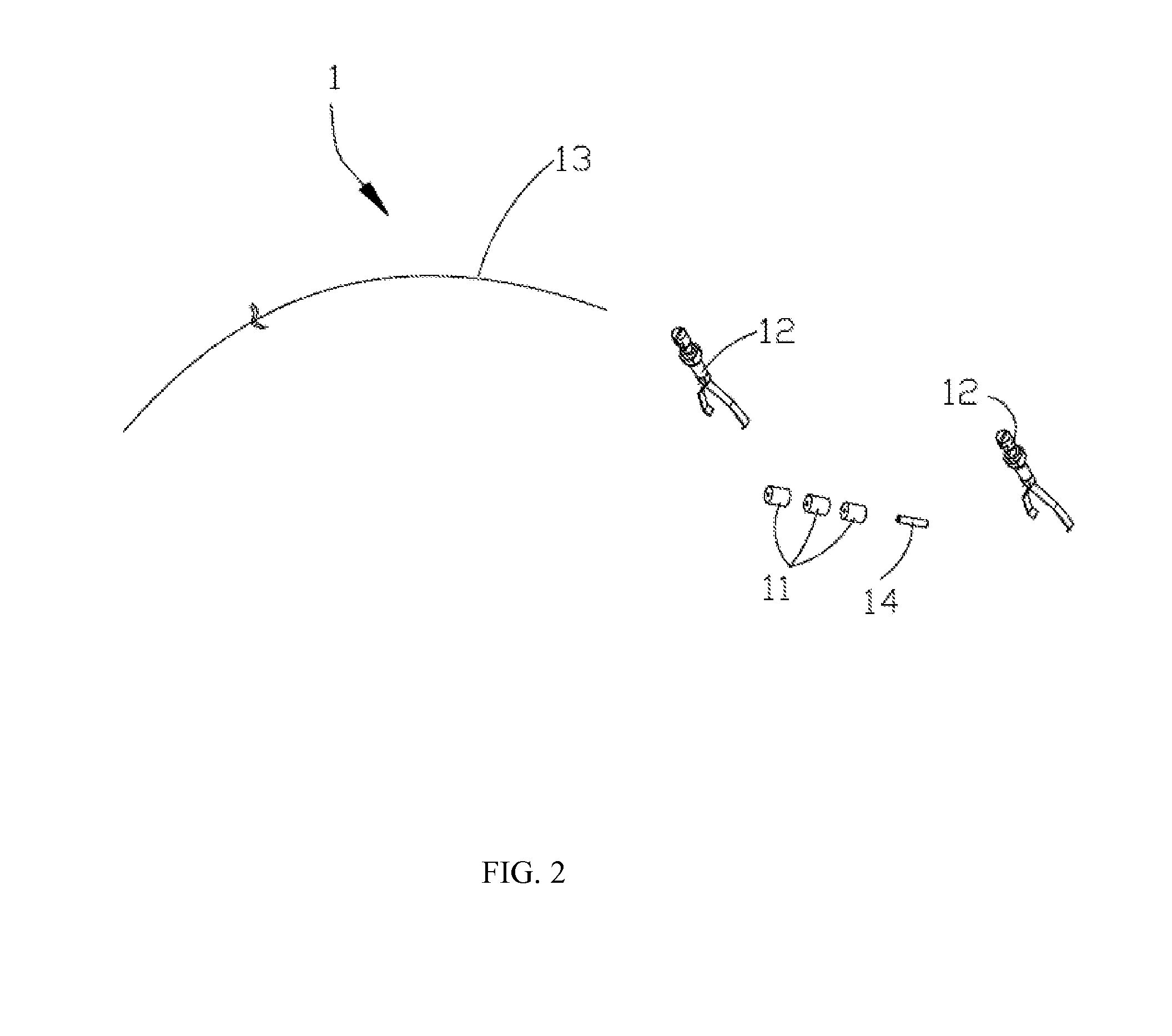
[0102]FIG. 7 to FIG. 10 schematically illustrate the operation process of the present disclosed auxiliary apparatus, which is uniformly radially polarized. Referring to FIG. 7, when an end of the surgery target 3 on a mucous membrane is resected, the clip 12 and the connective thread are delivered to appropriate site on the resected mucous membrane by the cannula-assisted endoscope 31. The clip 12, join to an end of the connective thread, takes hold of the surgery target 3 and fixes it at the incision of the mucous membrane. After that, the titanium clip releaser 33 separates the clip 12 from the cannula-assisted endoscope 31. FIG. 7 schematically illustrates the situation after the clip 12 is dismissed from the cannula-assisted endoscope 31.

[0103]FIG. 8 schematically illustrates the in vivo magnet 11 and the holding set 14 being introduced into the body cavity. Assisted by the guiding wire 32, the cannula-assisted endoscope 31 sequentially disposes the in vivo magnet 11, which is c...

second embodiment

[0108]FIG. 11 to FIG. 12 schematically illustrate the operation process of the present disclosed auxiliary apparatus, which is uniformly longitudinally polarized. As depicted in FIG. 10, after the surgery target 3 is resected, the clip 12 and the connective thread are delivered to the appropriate site on the resected mucous membrane by the cannula-assisted endoscope 31. The clip 12, joined to one end of the connective thread, anchors to the incision of mucous membrane, and then is released from the cannula-assisted endoscope 31.

[0109]After that, as illustrated in the first embodiment, the in vivo magnet 11 and the holding set 14 are delivered into the body cavity. With the assistance of the guiding wire 32, the cannula-assisted endoscope 31 sequentially threads the in vivo magnet 11, which is constructed of four magnetic cylinders having hollow column-shaped centers, and a silicone tube on the connective thread, and then positions them to one side of the first clip 12 via the connec...

third embodiment

[0120]As depicted in FIG. 15 and FIG. 16, the first knot is tied at one end of the stiff connector 13. Then the stiff connector 13 sequentially passes through the first set of the in vivo magnet 11, the holding set 14 and the second set of the in vivo magnet 11. The second knot is tied at the other end of the stiff connector 13, close to the second set of the in vivo magnet 11. the sizes of the first and second knots match with the internal diameter of the in vivo magnets 11 to assemble two sets of the in vivo magnets 11 and the holding set 14 into an assembly.

[0121]In an embodiment, each set of the in vivo magnet 11 may couple with a supporting set 15 to form individual assembly. Alternatively, the stiff connector may be set in shape of cylinder, which can sequentially assist the first set of the in vivo magnets 11, the holding set 14 and the second set of the in vivo magnet 11 to house the supporting column 1512. In this instance, only one supporting set 15 is needed.

[0122]After t...


PUM


Abstract

An auxiliary apparatus for MIS including an in vivo device and an in vitro device to stretch a surgery target is provided. The in vitro device comprises an in vitro magnet field-generating element. The in vivo device comprises an in vivo magnet-anchoring element to the surgery target. The in vivo magnet moves and/or rotates according to the direction change of the external magnetic field, which is generated by the in vitro magnet field-generating element, and thereby the surgery target can move in a controlled speed and/or rotate in a controlled angle according to the direction change of the external magnetic field. A method to control the auxiliary apparatus is also provided.

Description

BACKGROUND[0001]1. Technical Field[0002]The present disclosure relates to an auxiliary apparatus for minimally invasive surgery, and, more particularly, for assisting pulling or cutting off a target lesion area in a surgery.[0003]2. Description of Related Art[0004]Minimally invasive surgery (MIS) utilizes medical scopes, such as gastroscope, enteroscopy, laparoscopy, thoracoscope and relative tools to perform a surgery. The concept of minimal invasion derives from the development of the whole medical pattern and the idea of entire treatment. MIS focuses on the progress and recovery of patients' psychology, sociality, physical pain, spirit and life quality. It takes care of patients and reduces their suffering. During an MIS procedure, surgery targets are accessed through natural orifices or from one to three small incisions 0.5 cm to 1 cm in length, rather than through a large incision in an open surgery approach. The advantages of MIS over conventional surgeries include minimal sca...

Claims


Application Information

Patent Timeline
Patent Type & Authority Applications(United States)
IPC IPC(8): A61B17/02A61B17/00
CPCA61B17/0218A61B2017/00269A61B2017/00876A61B17/00234A61B17/122A61B2017/00477A61B34/73A61B1/00158A61B1/041A61L31/022
Inventor DUAN, XIAODONGZHANG, SHAOBANG
Owner ANKON MEDICAL TECH (SHANGHAI) CO LTD
Why Eureka
Try Eureka