
Multi-photon absorption for femtosecond micromachining and refractive index modification of tissues

a multi-photon absorption and tissue technology, applied in the field of vision correction, can solve the problems of tissue destruction, high cost of femtosecond laser flap cutting, and limited tissue destruction,

Active Publication Date: 2017-06-08
UNIVERSITY OF ROCHESTER
View PDF0 Cites 11 Cited by
  • Summary
  • Abstract
  • Description
  • Claims
  • Application Information

AI Technical Summary

Benefits of technology

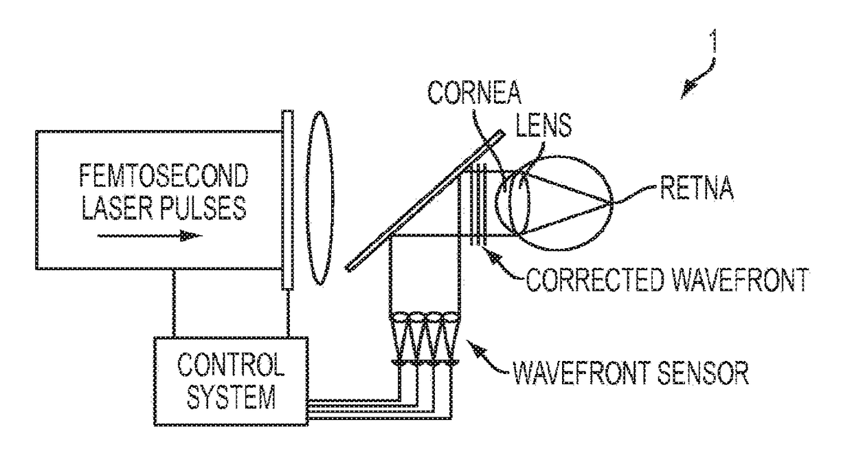
[0089]Ongoing experiments in our laboratories are generating information about the cellular and molecular mechanisms underlying IRIS in the living cornea, and allowing us to gain critical knowledge that can be used to further manipulate the size, placement and design of micromachined patterns, as well as the magnitude of the refractive index changes with which they are associated. The ability to alter the native refractive index of the cornea and lens without causing significant tissue damage has important theoretical and practical implications. By understanding how laser power can be used to alter tissues non-destructively, and by understanding the nature of these alterations, we could open up an entirely new branch in the field of laser biology. Among other things, this could completely change our approach to laser refractive surgery, and to vision correction more generally. For instance, the preservation of tissue clarity during the treatment allows the application of IRIS for the creation of corneal fiducial markings that could be used to align eye trackers during LASIK, and for refractive corrections in a closed-loop approach, e.g. with specific benefit for the correction of higher-order aberrations, as well as for “touch-up corrections” of ocular surface defects. More broadly, the feasibility of IRIS in living tissues would offer new possibilities for non-invasive alterations, marking or pattern-inscription within living organisms. From a theoretical stand-point, it also provides a unique opportunity to better understand and define the extent to which we can optically manipulate even large areas of living tissues without inducing a significant wound healing reaction.

Problems solved by technology

While increasingly popular, femtosecond laser flap cutting remains limited by its high cost, accessibility, as well as uncertainty about its long-term photochemical, mechanical and biological effects (Netto, Mohan, Medeiros, Dupps, Sinha, Krucger, Stapleton, Rayborn.
Recent reports have detailed negative side-effects of this technique, particularly in terms of tissue destruction, which, at some laser settings, appears stronger than following mechanical microkeratome cuts (Netto et al., 2007, Stonecipher et al., 2006).
As a result, absorbed energy accumulates locally, with destructive consequences (Schaff, Garcia & Mazur, 2003).
However, whether threshold fluence increases or decreases as pulse durations get shorter remains a challenging question (Stern et al., 1989, Stuart et al., 1995, Tien et al., 1999).
Some models predict that the threshold would first increase, then decrease when pulse duration becomes shorter than 100 fs, but there is no solid experimental evidence to support this (Tien et al., 1999).
However, insufficient experimentation on cornea and lens using sub-100 fs pulses makes it difficult to support this prediction and furthermore, existing data were collected using single pulses from low-repetition-rate lasers.
Advantages and disadvantages of the femtosecond laser microkeratome.

Method used


Image

  • Multi-photon absorption for femtosecond micromachining and refractive index modification of tissues
  • Multi-photon absorption for femtosecond micromachining and refractive index modification of tissues
  • Multi-photon absorption for femtosecond micromachining and refractive index modification of tissues

Examples


Embodiment Construction

[0108]A preferred embodiment of the present invention will now be set forth in detail with reference to the drawings.

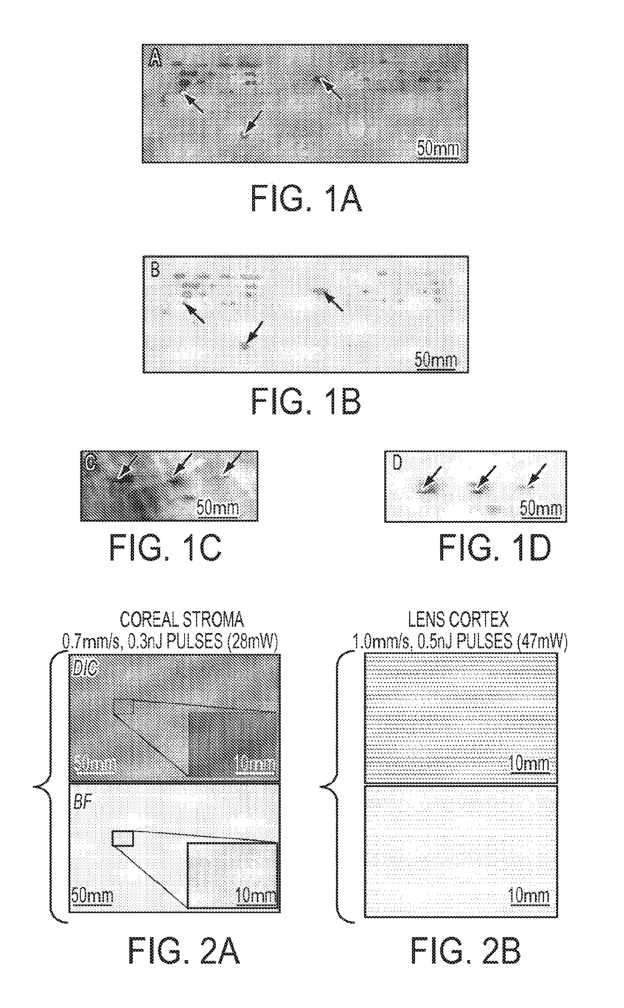
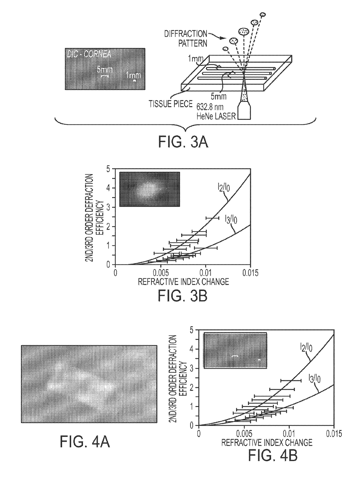
[0109]Our preliminary experiments (Ding, Huxlin & Knox, 2007, Ding et al., 2008, Huxlin, Ding & Knox, 2008) showed that it is possible to change the refractive index of the lightly-fixed, mammalian cornea and lens without tissue destruction, a phenomenon we termed Intra-tissue Refractive Index Shaping (IRIS). To achieve this, we first measured, then reduced femtosecond laser pulse energies below the optical breakdown threshold of lightly-fixed post-mortem cat corneas and lenses. In both silicone and non-silicone-based hydrogels, this approach induced a significant change in refractive index without visible plasma luminescence or bubble formation (Ding et al., 2006).

[0110]Eight corneas and eight lenses were extracted under surgical anesthesia from five normal, adult domestic short-hair cats (felis cattus). To avoid decomposition and opacification prior to femtosecond l...


PUM


Abstract

By adapting femtosecond micromachining approaches developed in hydrogels, we can perform Intra-tissue Refractive Index Shaping (IRIS) in biological tissues. We reduced femtosecond laser pulse energies below the optical breakdown thresholds to create grating patterns that are associated with a change in the refractive index of the tissue. To increase two-photon absorption, we used a two (or more)-photon-absorbing chromophore.

Description

REFERENCE TO RELATED APPLICATIONS[0001]The present application is a continuation of U.S. patent application Ser. No. 12 / 207,137, filed Sep. 9, 2008, currently pending, which is a continuation-in-part of U.S. patent application Ser. No. 12 / 146,976, filed Jun. 26, 2008, now U.S. Pat. No. 8,486,055, which claims the benefit of U.S. Provisional Patent Application Nos. 60 / 929,397, filed Jun. 26, 2007, and 61 / 026,890, filed Feb. 7, 2008. The disclosures of all of those applications are hereby incorporated by reference in their entireties into the present application.STATEMENT OF GOVERNMENT INTEREST[0002]This invention was made with government support under Grant Nos. EY015836 and EY001319 awarded by the National Institutes of Health. The government has certain rights in the invention.FIELD OF THE INVENTION[0003]The present invention is directed to vision correction and more particularly to vision correction by modifying the index of refraction of ocular tissue in the eye by a high-repetit...

Claims


Application Information

Patent Timeline
Patent Type & Authority Applications(United States)
IPC IPC(8): A61F9/008
CPCA61F9/00827A61F9/00804A61F9/00829A61F2009/00872A61F2009/00848A61F2009/0087A61F2009/00842A61F9/008A61F2009/0088A61F2009/00846
Inventor KNOX, WAYNE H.HUXLIN, KRYSTEL R.DING, LI
Owner UNIVERSITY OF ROCHESTER
Learn More