
Molecular Display System

a molecular display and molecular target technology, applied in the field of molecular display methods, can solve the problems of poor enrichment of cell surface antigens, difficult selection of libraries against cell-surface antigens, and inability to obtain certain epitopes for binding, etc., and achieve the effect of rapid and efficien

Active Publication Date: 2017-06-29
THE GOVERNING COUNCIL OF THE UNIV OF TORONTO
View PDF0 Cites 1 Cited by
  • Summary
  • Abstract
  • Description
  • Claims
  • Application Information

AI Technical Summary

Benefits of technology

The patent describes a method for identifying and recovering binding agents, such as antibodies, that specifically target cell-surface targets. The method involves screening a library of potential binding agents, sequencing the genetically encoded affinity reagents, and identifying the sequences that are more abundant in the positive selection pool. This method does not require additional purification or isolation steps prior to identification and recovery of the binding agent. The patent also describes the use of deep sequencing and recovery strategies, as well as differential selection strategies to identify and select specific binding agents. The methods can be used with phage-display libraries and can be used in combination with other peptide / polypeptide display systems. The patent also provides antibodies and antibody fragments that specifically target cell-surface targets, including CDR regions. These antibodies can be used for the treatment of cancer, among others.

Problems solved by technology

For example, phage display antibody technologies are used for isolating antibody fragments specific to antigens of interest, but selection of libraries against cell-surface antigens remains very challenging.
Consequently, poor enrichment for binding phage clones is typically observed in cell selections.
If a protein is not properly folded, certain epitopes may not be available for binding by, for example, an affinity reagent.
Likewise, proteins that are part of large complexes or associated with DNA, histones or other subcellular structures contain epitopes that are not necessarily made available for binding following traditional purification methods.
For example, the properties of multi-pass membrane G-protein coupled receptors make their expression and purification very difficult, yet they are particularly relevant drug targets [1,2].
Although these methods may help to enrich for phage clones specific to the antigen of interest, the number of unique antibody fragments recovered by these methods often remains relatively low, as phage display methodologies typically exhibit an affinity based selection pressure that promotes sequence convergence in later rounds of selection.

Method used


Image

  • Molecular Display System
  • Molecular Display System
  • Molecular Display System

Examples


example 1

Rapid Isolation of Antibody Fragments to Cell-Surface Targets

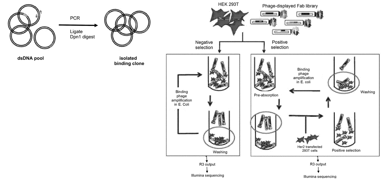
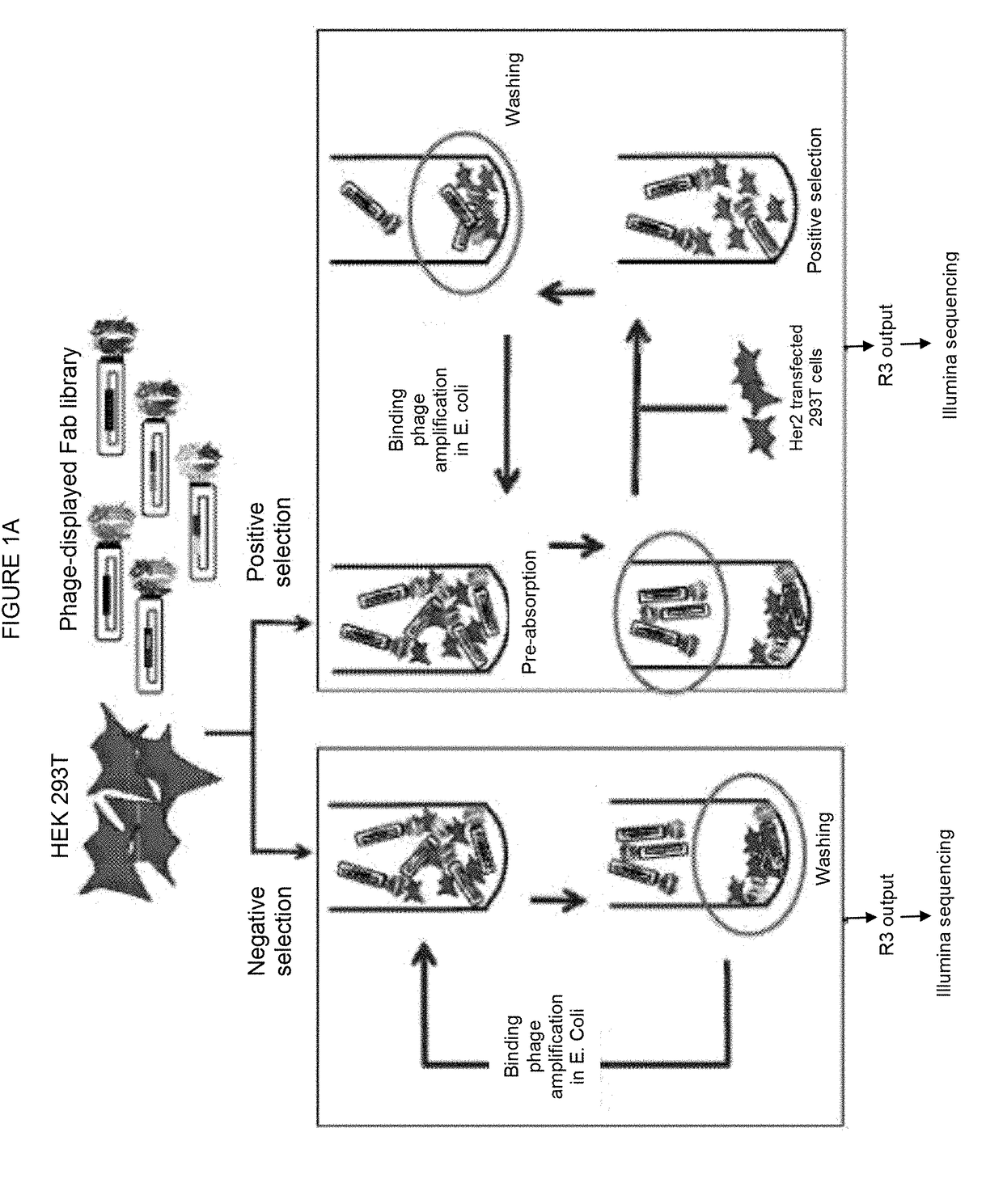
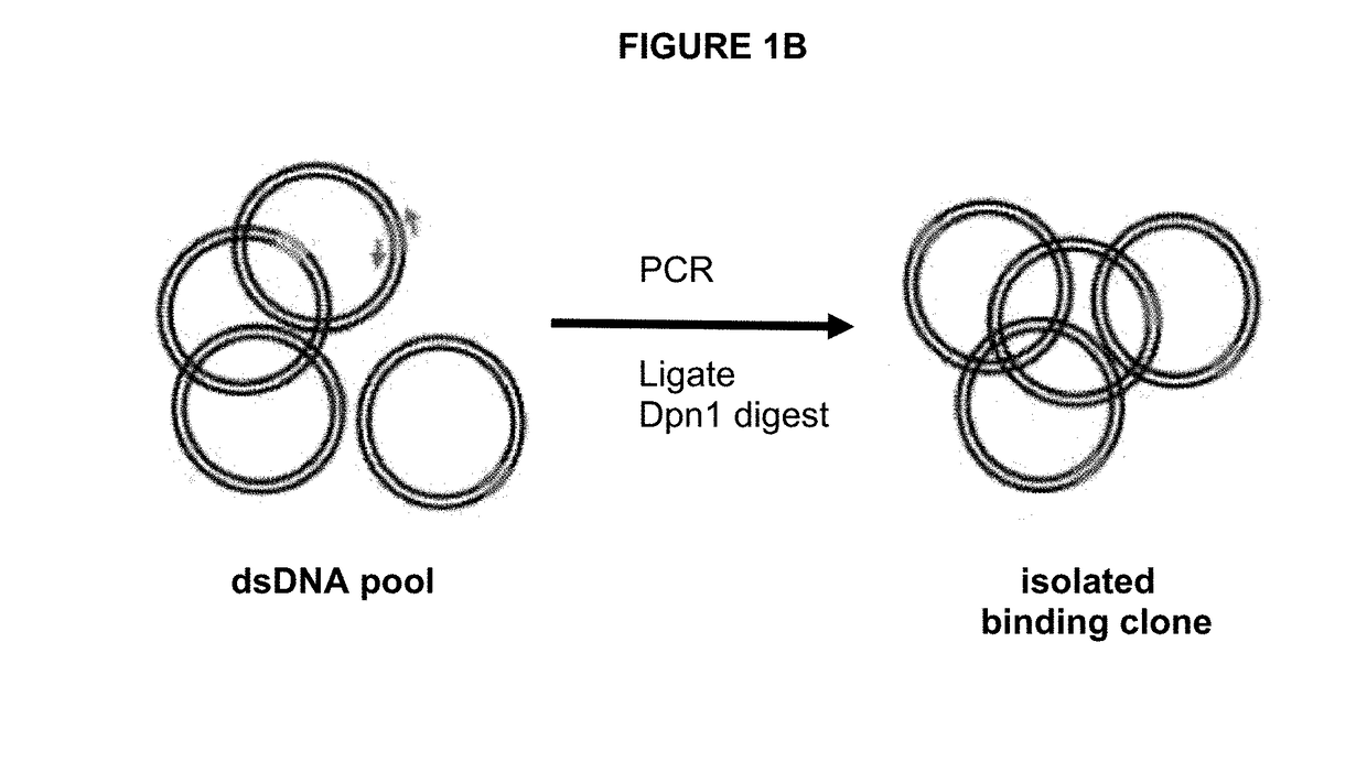
[0070]The considerable heterogeneity of cell-surfaces makes selection of phage-displayed antibody libraries against cell-surface antigens challenging. We report the development of a unique methodology for rapidly isolating phage-displayed antibody fragments to cell-surface targets, using the oncogenic human epidermal growth factor receptor 2 (Her2) as a model. Synthetic phage-displayed libraries were selected in parallel on Her2-positive and negative cells. Following three rounds of selection, the output phage pools were analyzed by Illumina deep sequencing. Comparisons of the sequences from the positive and negative selection pool allowed sequences specific to the antigen-expressing cell-line to be readily identified from background phage clones. A PCR amplification strategy that used primers specific to the unique heavy chain third hypervariable loop enabled the recovery of clones from the positive selection pool, which ...

example 2

PCR-Based Recovery of Antibody Fragments to Additional Cell-Surface Targets

[0098]The rescue strategies described herein make use of both the unique H3 and L3 CDR sequences.

[0099]As an alternative to identifying positive Fabs by clonal cell ELISAs, two different PCR based recovery methods are used (FIGS. 6A, 6B). As depicted in FIG. 6A, two primer sets specific for both CDR H3 and L3 are used to make the recovery more specific. Primers are designed to anneal to the L3 and H3, and amplify two fragments, in both directions. This results in two fragments that both contain the L3 and H3 regions. The two fragments can be annealed, and then a single round of DNA extension is done. The resulting product can then be ligated and transformed into E. coli to recover the desired phage clone in the original library display vector. As depicted in FIG. 6B, three primer sets are used to amplify three fragments, in a strategy that makes use of both the H3 and L3 unique sequences and unique Nsi1 and N...

example 3

Rapid Isolation of Antibody Fragments Specific to Cell Growth Conditions

[0101]FIG. 7 depicts a flow chart of the selection strategy used to isolate Fab clones specific for cell surface O-GlcNAc-dependent epitopes, demonstrating that the CellectSeq method can be used to isolate Fabs specific to cell growth conditions. The positive selection begins a pre-absorption step in which the library phage are incubated with MCF7 breast cancer cells grown in DMEM (Dulbecco's Modified Eagle Medium) (high glucose version) supplemented with 10% FBS. After incubation, the mixture is pelleted to remove the library clones bound to the cells. These clones are likely specific for cell-surface epitopes that are not of interest, or are non-specific binding clones. The library phage remaining in the supernatant are incubated with the MCF7 cells grown in DMEM (high glucose version) supplemented with 10% FBS plus 30 mM Glucosamine (G), 5 mM Uridine (U) and 50 μM PugNAc (P) (collectively referred to as GUP),...


PUM

PropertyMeasurementUnit
temperatureaaaaaaaaaa
flow rateaaaaaaaaaa
surfaceaaaaaaaaaa

Abstract

There is provided herein a method for identifying and / or recovering at least one genetically encoded affinity reagent specific for a target molecule by screening using molecular display in conjunction with the sequencing of positive and negative selection pools from the screen.

Description

RELATED APPLICATIONS[0001]This application is a continuation of U.S. patent application Ser. No. 13 / 629,520, filed Sep. 27, 2012, now abandoned, which claims the benefit of provisional application U.S. Ser. No. 61 / 539,546 filed Sep. 27, 2011, the contents of which are herein incorporated by reference in their entirety.INCORPORATION OF SEQUENCE LISTING[0002]The contents of the text file named “UTOR004C01US_SubSL.txt”, which was created on Jan. 18, 2017 and is 73.8 MB in size, are hereby incorporated by reference in their entirety.FIELD OF INVENTION[0003]This invention relates to the field of screening for affinity reagents to a molecular target, and more specifically to molecular display methods used in conjunction with sequencing.BACKGROUND[0004]Molecular display technologies are widely used to screen for potential affinity binders to a specific target molecule, however, there is potential for improving thereon. For example, phage display antibody technologies are used for isolating...

Claims


Application Information

Patent Timeline
Patent Type & Authority Applications(United States)
IPC IPC(8): G01N33/68
CPCG01N33/6857G01N33/6854C12N15/1037C40B30/04
Inventor SIDHU, SACHDEVMOFFAT, JASONPERSSON, HELENAPATEL, NISHSUNDARARAJAN, SARAVANANGAKHAL, AMANDEEPYE, WEITOMIC, JELENAMCLAUGHLIN, MEGAN ELIZABETH
Owner THE GOVERNING COUNCIL OF THE UNIV OF TORONTO
Learn More