Method for gestational age estimation and embryonic mutant detection

a technology of gestational age estimation and mutant detection, applied in the field of streamlining development biology studies, can solve the problems of inaccuracy of method, inconsistent cross sectional area, and difficulty in image segmentation of bvs in hfu or other chosen imaging modality, and achieve the effect of accurate staging of embryos

Active Publication Date: 2017-07-27
RIVERSIDE RES INST +1
View PDF0 Cites 4 Cited by
  • Summary
  • Abstract
  • Description
  • Claims
  • Application Information

AI Technical Summary

Benefits of technology

[0006]The present invention relates to characterizing the shape variation of the BVs during embryonic growth and due to genetic mutations. The present invention provides a framework to characterize the shape variation of BVs by extracting the skeleton of the BV region, and decomposing the BV region into five components automatically based on the volume profile along the skel

Problems solved by technology

Image segmentation of the BVs in HFU or other chosen imaging modality can be difficult and time consuming, particularly if some portions of boundaries are missing.
However, the cross sectio

Method used


Image

  • Method for gestational age estimation and embryonic mutant detection
  • Method for gestational age estimation and embryonic mutant detection
  • Method for gestational age estimation and embryonic mutant detection

Examples


example

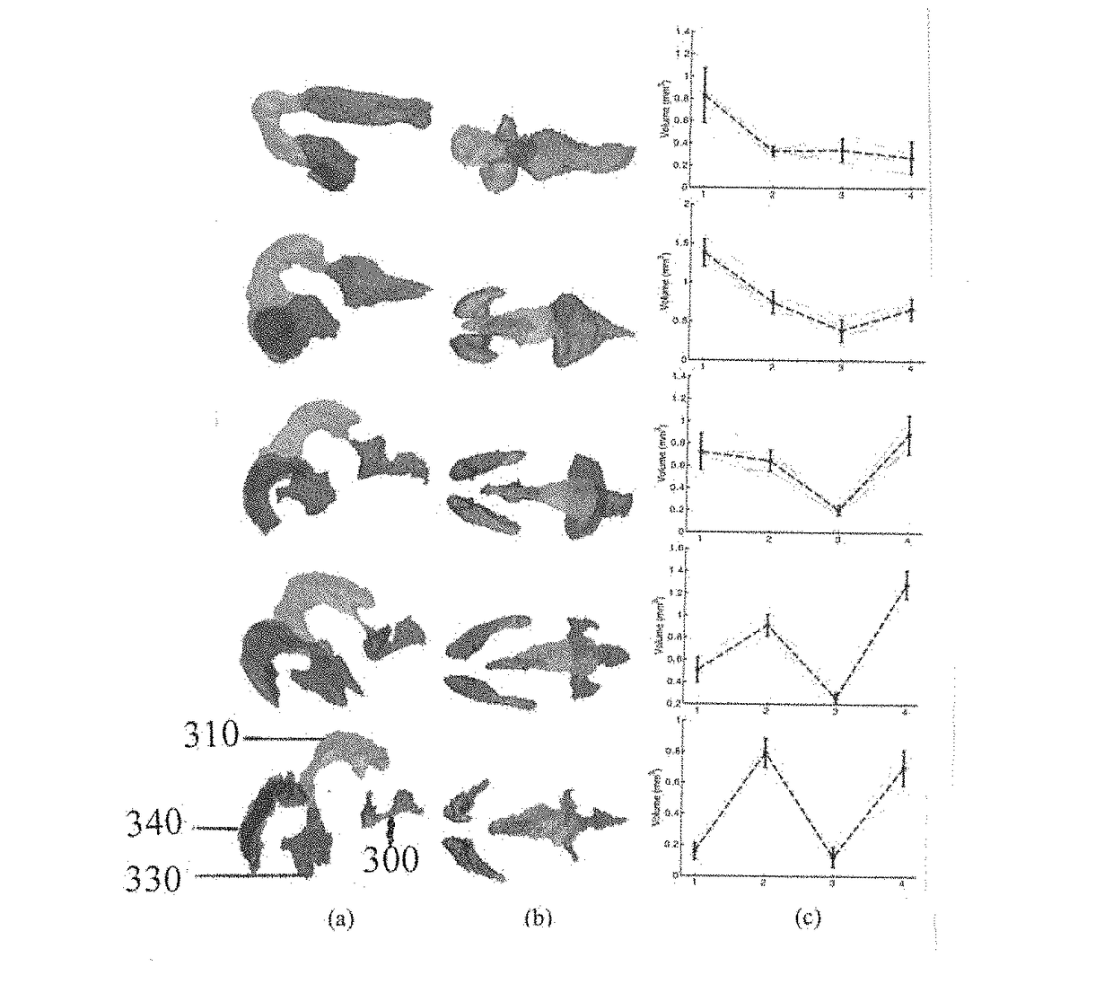
[0034]Volumetric ultrasound data were acquired in utero and in vivo from pregnant mice using a 5-element, 40-MHz annular array. A 5-channel pulser was used to consecutively excite each array element. The 25 resulting transmit / receive signals processed using delay-and-sum beamforming yielded a depth of field sufficient to cover the entire head of the embryo. The method was applied to 40 wild type embryos spanning from days E10.5 to E14.5.

[0035]A. Staging

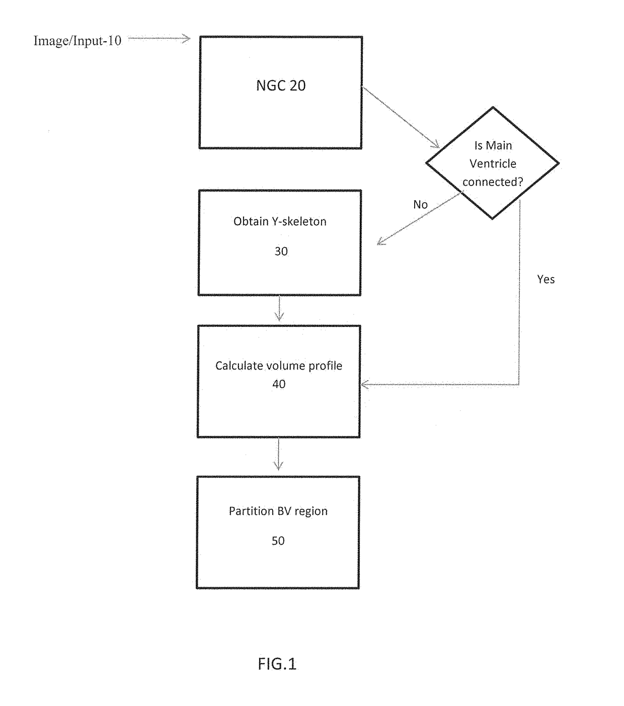
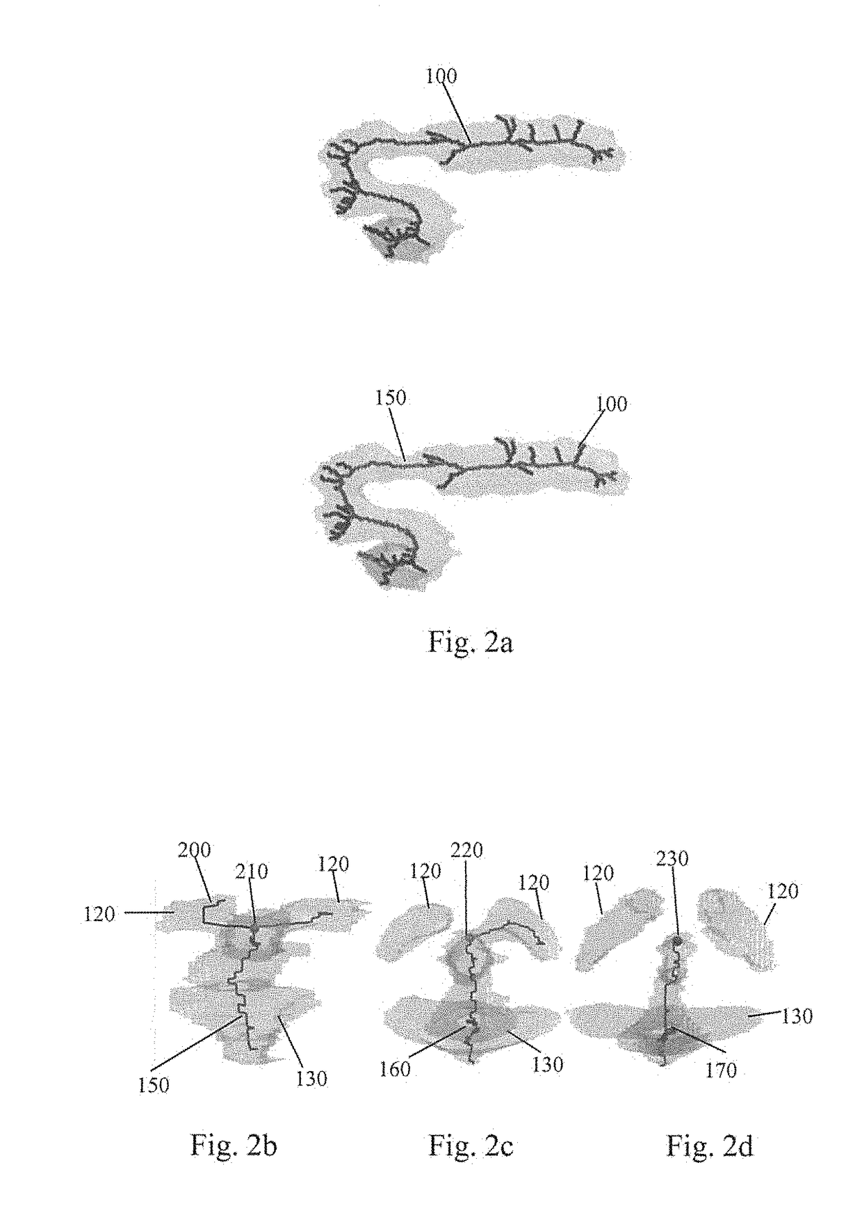
[0036]The method was tested on 40 mouse embryo head-region images crossing five gestation stages. NGC was used to segment the BVs in each image and its Y-skeleton and the volume profile along the skeleton were derived from the segmentation of the images. Based on the volume profile, the BV region was partitioned into five components and the volume vector was calculated. FIGS. 3a-3c show the partitioning results for a typical image from each gestation stage, the volume vectors computed for all images and the mean volume vector for each...


PUM


Abstract

A method to characterize shape variations in brain ventricles during embryonic growth in mammals, the method including extracting a brain ventricle skeleton from one or more images, calculating a volume profile for the skeleton using the extracted images, partitioning the brain ventricle based on the volume profile along the skeleton, the brain ventricle being partitioned into two lateral ventricles and a main ventricle, the main ventricle being further partitioned into three sub regions, determining volume vectors of the two lateral ventricles and the three sub regions, computing a means square error between the determined computed volume vectors and a pretrained mean volume vector of embryos during different gestational stages, and classifying the embryo to the gestational stage having the lowest mean square error. A method to characterize mutant detection in mammals, the method including acquiring one or more images, computing a volume profile directly along a path of the detected skeleton from the one or more images, aligning the volume profile against a standard profile, and evaluating the volume profile against the standard profile to detect a mutation.

Description

PRIORITY AND RELATED APPLICATION[0001]This application claims priority to U.S. Provisional Patent Application No. 62 / 281,284 filed Jan. 21, 2016 entitled “METHOD FOR GESTATIONAL AGE ESTIMATION AND EMBRYONIC MUTANT DETECTION” and is hereby incorporated by reference in its entirety.FIELD OF THE INVENTION[0002]The present invention relates to streamlining development biology studies. More specifically the present invention relates to gestational staging and mutant detection.BACKGROUND OF THE INVENTION[0003]The analysis of brain ventricles (BVs) is important in the study of normal and abnormal development of the central nervous system. The mouse has been used as a model to study mammalian development because of the high degree of homology between the mouse and human genome. Studying how mutations manifest themselves during embryonic development in mice assists in the study of the human central nervous system. Real time imaging modalities and automated image processing methods are needed...

Claims


Application Information

Patent Timeline
IPC IPC(8): G06T7/00
CPCG06T7/0012G06T2207/30016G06T2207/30044G06T2207/10136
Inventor KUO, JEN-WEIMAMOU, JONATHANKETTERLING, JEFFREY A.ARISTIZABAL, ORLANDOTURNBULL, DANIEL H.WANG, YAO
Owner RIVERSIDE RES INST
Why Eureka
Try Eureka