
Method and device for image guided post-nasal nerve ablation

a post-nasal nerve and image-guided technology, applied in the field of cryosurgical probes, can solve the problems of increasing the infection rate, affecting the quality of life, and removing or ablation of the mucosal tissue including the surface epithelial layer, so as to reduce or interrupt the rhinitis, the effect of reducing or eliminating the rhinitis

Active Publication Date: 2017-08-17
ARRINEX INC
View PDF10 Cites 32 Cited by
  • Summary
  • Abstract
  • Description
  • Claims
  • Application Information

AI Technical Summary

Benefits of technology

The patent describes a device and method for reducing or eliminating nasal rhinitis by ablating certain nerve bundles. The device includes an imaging assembly that can be articulated to a desired viewing angle or position relative to a therapeutic element, which can also be articulated. The imaging assembly is designed to minimize engagement with nasal tissue, and the therapeutic element can be positioned adjacent to the lateral nasal wall tissue region for improved therapeutic effects. The technical effects of this patent include improved visualization and reduced invasiveness of using the device, as well as improved therapeutic outcomes.

Problems solved by technology

In particular, chronic severe nasal obstruction resulting from remodeling of submucosal tissues of the inferior turbinates due to dilation of the venous sinuses or fibrosis can interfere with the quality of life (QOL).
Removal or ablation of the mucosal tissue including the surface epithelial layer has the disadvantage of postoperative complications such as crusting and an increased infection rate.
Turbinectomy is thought to have the greatest effect on nasal obstruction, and slight improvement in some rhinitis patients but it is accompanied by severe adverse effects such as bleeding, crusting, and nasal dryness.
However, the original technique was abandoned at the beginning of the 1980s because of its irreversible complications such as dry eyes.
However, it can be difficult to deliver energy to the correct location without direct or indirect visualization.
Such visualization devices are expensive, bulky, and difficult to operate simultaneously with energy delivery devices.
For example, using an energy delivery device with a rigid endoscope may require the healthcare provider to use both hands, or may require a second individual to perform the procedure, which may make the procedure more time consuming and costly.
Moreover, separate rigid or flexible endoscopes and existing visualization devices may not allow a healthcare provider to access far enough into the target anatomy.

Method used


Image

  • Method and device for image guided post-nasal nerve ablation
  • Method and device for image guided post-nasal nerve ablation
  • Method and device for image guided post-nasal nerve ablation

Examples


Embodiment Construction

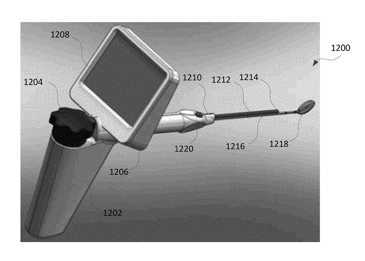
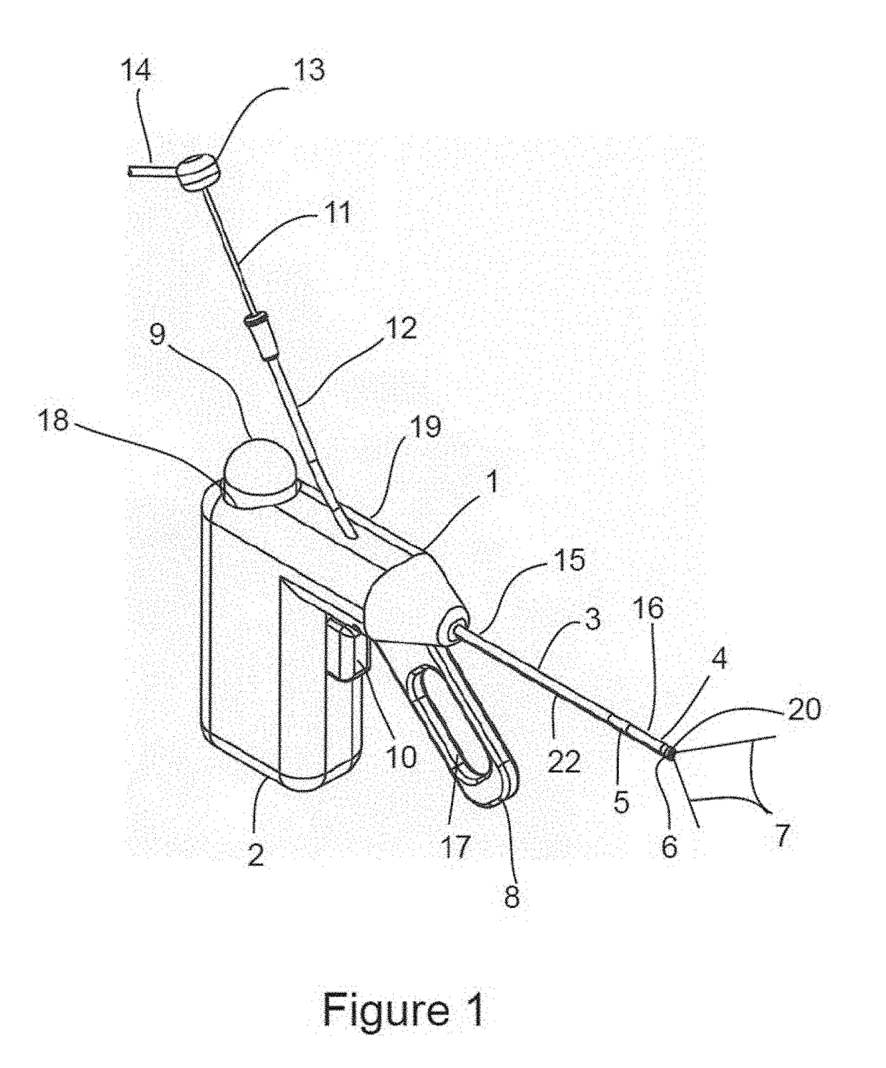
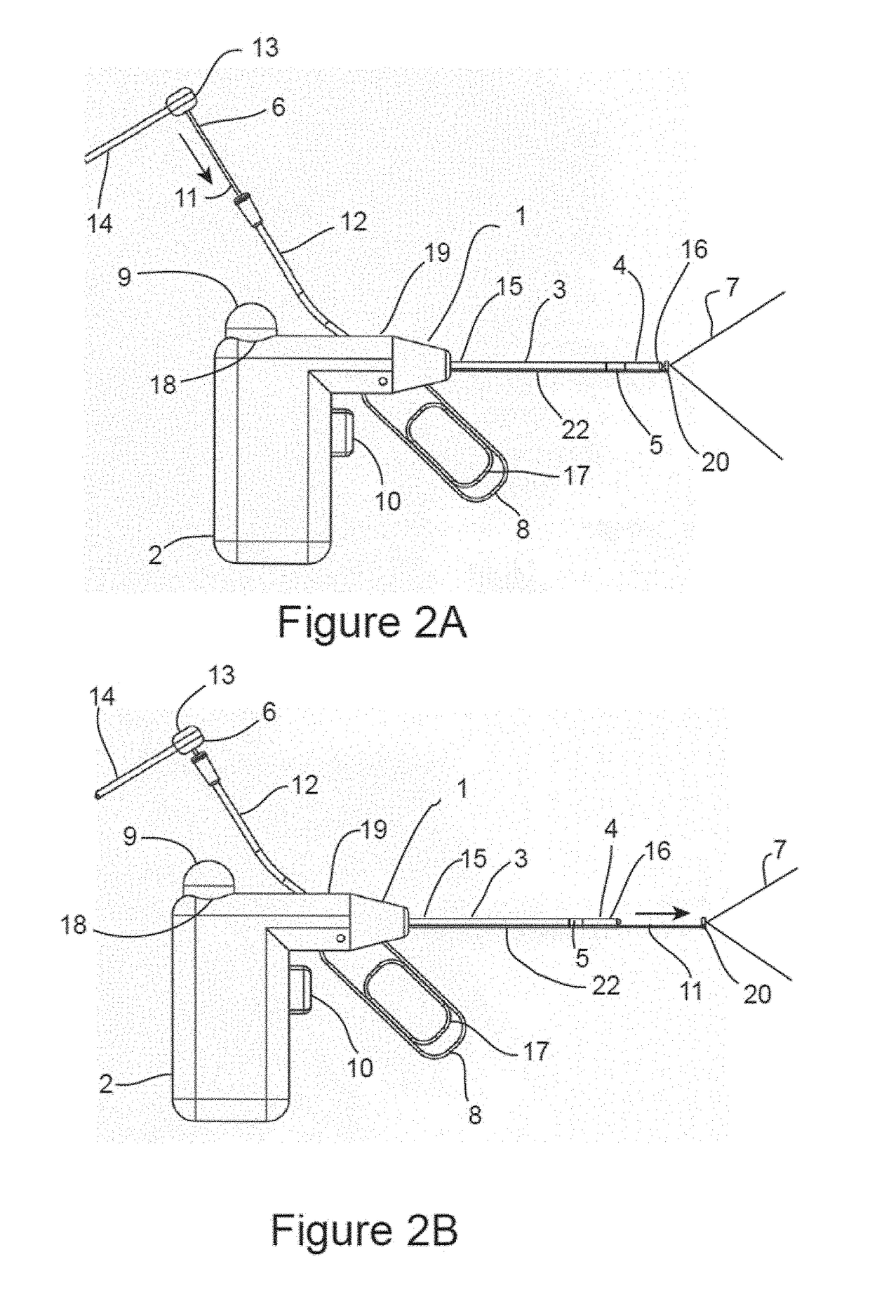
[0066]FIG. 1 is a schematic illustration of surgical ablation probe 1 configured for ablation of posterior nasal nerve function. As depicted in the figures, surgical ablation probe 1, and its alternative embodiments are cryo-ablation probes. However, alternative ablation and therapeutic modalities, including radiofrequency, laser, microwave, ultrasonic, and chemo-ablation remain within the scope of this invention. Surgical ablation probe 1 comprises handle assembly 2, probe shaft 3, and camera assembly 6. Handle assembly 2 comprises handle housing 19, cryogen cartridge receptacle 18, cryogen cartridge 9, cryogen control trigger 10, distal segment actuator lever 8 with finger grip 17, and camera tube 12. Probe shaft 3 comprises proximal end 15, distal end 16, cryo-ablation element 4, distal articulated segment 5, proximal segment 21, and camera channel 22. Camera assembly 6 comprises camera head 20, camera shaft 11, camera hub 13, camera electrical cable 14, and camera field of view ...


PUM


Abstract

Devices and methods for treating rhinitis are provided. An integrated therapy and imaging device is provided for single handheld use. The device may have a hollow elongated cannula, a therapeutic element coupled to a distal portion of the cannula, an imaging assembly coupled to the cannula to provide visualization of the therapeutic element, and an articulating region operably coupled to the imaging assembly to articulate the imaging assembly. The imaging assembly may be articulated so as to translate vertically, laterally, axially, and / or rotationally.

Description

FIELD OF THE INVENTION[0001]This application claims the benefit of U.S. Provisional Application No. 62 / 294,142, filed Feb. 11, 2016, which application is incorporated herein by reference in its entirety.[0002]The present invention relates to cryosurgical probes and their methods of use. More particularly, the present invention relates to cryosurgical probes which are configured to be advanced into a nasal cavity for treating conditions such as rhinitis.BACKGROUND OF THE INVENTION[0003]The major symptoms of allergic or non-allergic chronic rhinitis are sneezing, rhinorrhea, and night time coughing which are brought about by mucosal swelling, hyper-responsiveness of the sensory nerves, and an increased number and augmented responses of secretory cells in the inferior turbinates, respectively. In particular, chronic severe nasal obstruction resulting from remodeling of submucosal tissues of the inferior turbinates due to dilation of the venous sinuses or fibrosis can interfere with the...

Claims


Application Information

Patent Timeline
IPC IPC(8): A61B1/00A61B1/05A61B1/06A61B1/015A61B18/14A61B18/24A61B1/005A61B1/233A61B18/06
CPCA61B1/00087A61B2218/007A61B1/05A61B1/0676A61B1/00052A61B18/06A61B18/1492A61B18/24A61B1/00105A61B1/005A61B1/015A61B1/00066A61B1/00096A61B1/00098A61B1/00091A61B1/00094A61B2018/00327A61B2018/00577A61B2018/00982A61B2018/00994A61B2018/0212A61B2018/00434A61B2018/1861A61B2018/0022A61B2018/00714A61B2034/2057A61B2218/002A61B1/233A61B1/00082A61B1/0014A61B1/00181A61B1/0051A61B18/02A61B18/22A61B2090/3614A61B2090/309A61B90/361A61B1/0625A61B1/009
Inventor SAADAT, VAHIDSAADAT, MOJGANFOX, WILLIAM JASON
Owner ARRINEX INC
Features
  • Generate Ideas
  • Intellectual Property
  • Life Sciences
  • Materials
  • Tech Scout
  • Unparalleled Data Quality
  • Higher Quality Content
  • 60% Fewer Hallucinations
Learn More