Methods, Systems, and Apparatuses for Quantitative Analysis of Heterogeneous Biomarker Distribution

a biomarker and distribution technology, applied in image analysis, image enhancement, instruments, etc., can solve the problems of sample general deformation, deleterious effect, and not having a deleterious effect on processing

Active Publication Date: 2017-09-14
OREGON HEALTH & SCI UNIV OF PORTLAND OREGON +1
View PDF1 Cites 58 Cited by
  • Summary
  • Abstract
  • Description
  • Claims
  • Application Information

AI Technical Summary

Benefits of technology

[0044]In an exemplary embodiment, the foregoing system may further comprising a slide containing a cell sample in which one or more analytes of interest is labeled with a detectable label. In a further embodiment, the cell sample is a formalin-fixed paraffin embedded tissue sample that has been preserved using a two-temperature fixation, and said detectable label is attached to at least one antibody that specifically binds to at least one phosphorylated protein.

Problems solved by technology

Although increasing the diffusion time to 4 hours or greater generally had little beneficial effect, leaving tissues in the cold formalin for an extended period of time (for example, up to 14 days) generally does not have a deleterious effect on processing.
However, if the temperature is increased above about 50° C., the sample generally begins to degrade, which may have a deleterious effect on certain subsequent histological reactions.
Thus, relatively small changes in marker levels become difficult or impossible to discern.
To this data may be associated data from orthogonal studies such as sequencing or mass spec; keeping such data organized using conventional file formats and directory structures may become inefficient and cumbersome.
Dysregulation of these signaling pathways, whereby external stimuli may no longer be needed, can cause pathological aberration and the development of cancer.

Method used


Image

  • Methods, Systems, and Apparatuses for Quantitative Analysis of Heterogeneous Biomarker Distribution
  • Methods, Systems, and Apparatuses for Quantitative Analysis of Heterogeneous Biomarker Distribution
  • Methods, Systems, and Apparatuses for Quantitative Analysis of Heterogeneous Biomarker Distribution

Examples


Embodiment Construction

I. ABBREVIATIONS AND DEFINITIONS

[0068]In order to facilitate review of the various examples of this disclosure, the following explanations of abbreviations and specific terms are provided:

[0069]CISH: Chromogenic in situ hybridization.

[0070]CRC: colorectal cancer

[0071]FFPE tissue: Formalin-fixed, paraffin-embedded tissue.

[0072]FISH: Fluorescent in situ hybridization.

[0073]H&E: Hematoxylin and eosin staining.

[0074]IHC: Immunohistochemistry.

[0075]ISH: In situ hybridization.

[0076]NBF: neutral buffered formalin solution.

[0077]NSCLC: non-small cell lung cancer

[0078]PI3Ks: phosphatidylinositol 3-kinases. Also referred to as Phosphatidylinositol-4,5-bisphosphate 3-kinase, phosphatidylinositide 3-kinases, PI 3-kinases, PI(3)Ks, and PI-3Ks.

[0079]TNBC: triple negative breast cancer

[0080]Analyte: A molecule or group of molecules that are to be specifically detected in a sample.

[0081]Analyte-binding entity: Anything that is capable of specifically binding to an analyte. Examples of analyte-bindi...


PUM


Abstract

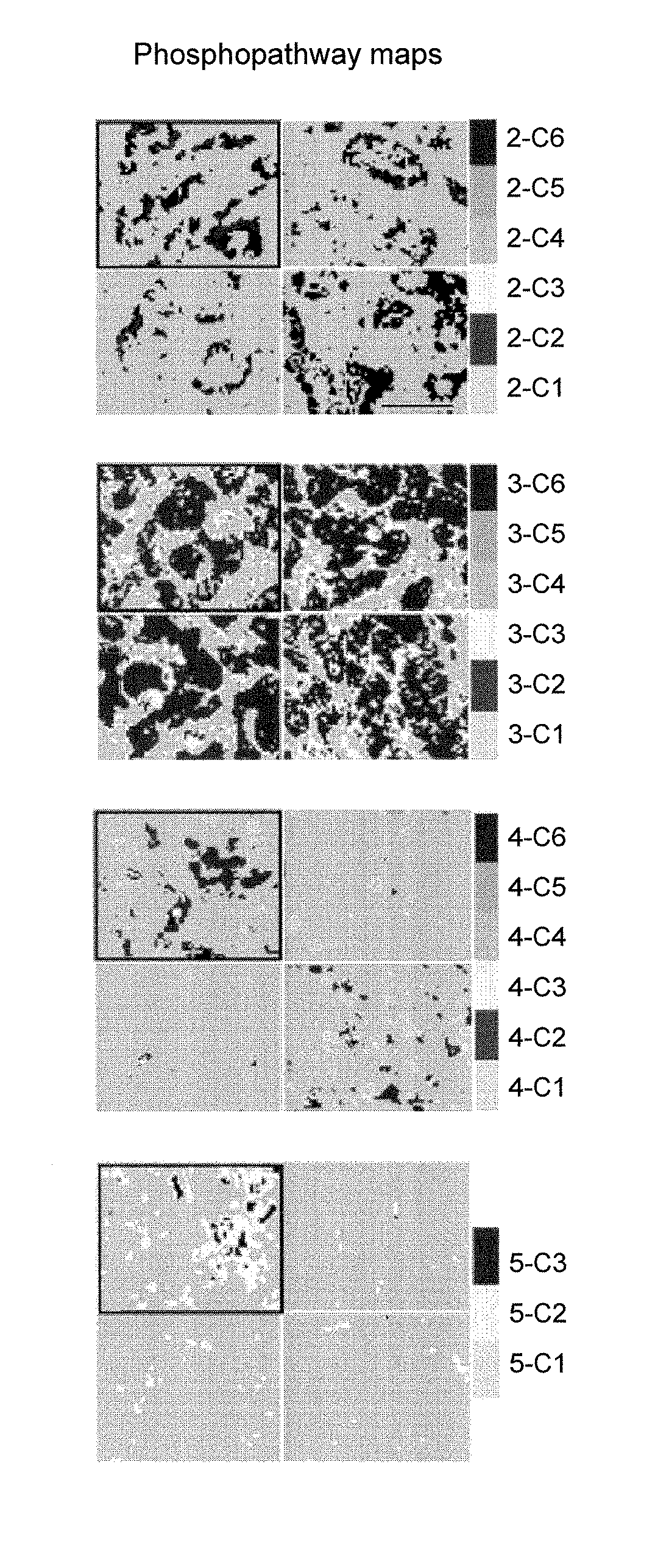
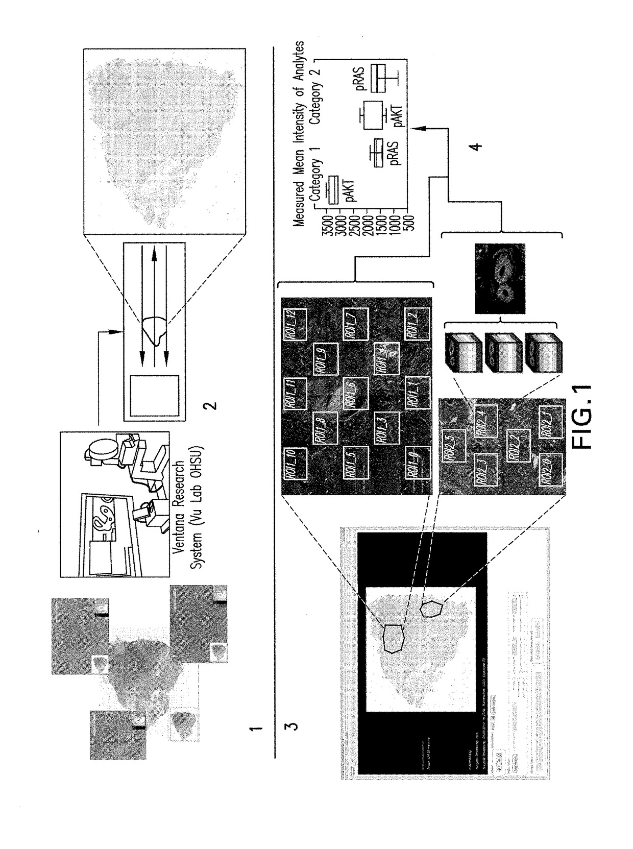
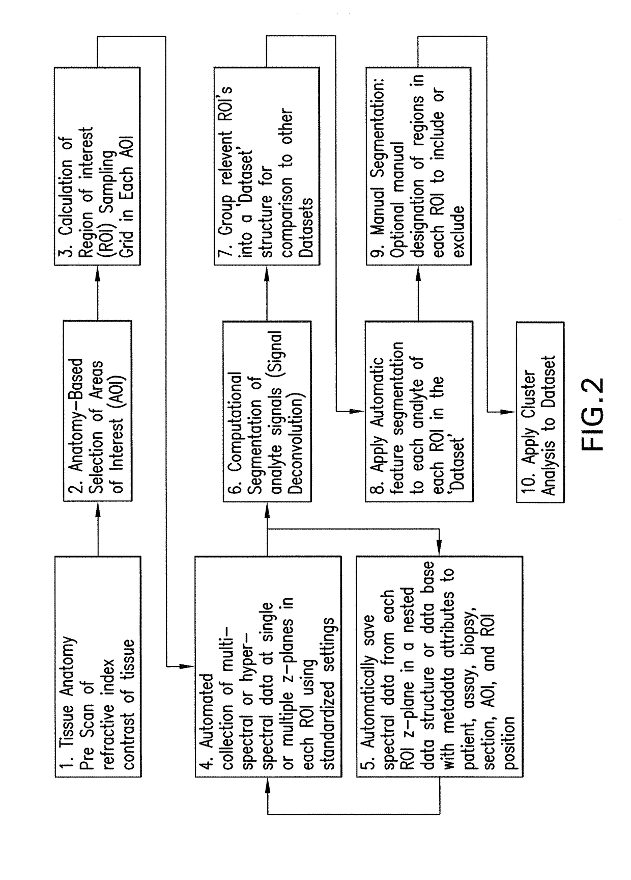
Methods, systems, and apparatuses for detecting and describing heterogeneity in a cell sample are disclosed herein. A plurality of fields of view (FOV) are generated for one or more areas of interest (AOI) within an image of the cell sample are generated. Hyperspectral or multispectral data from each FOV is organized into an image stack containing one or more z-layers, with each z-layer containing intensity data for a single marker at each pixel in the FOV. A cluster analysis is applied to the image stacks, wherein the clustering algorithm groups pixels having a similar ratio of detectable marker intensity across layers of the z-axis, thereby generating a plurality of clusters having similar expression patterns.

Description

CROSS-REFERENCE TO RELATED APPLICATIONS[0001]This patent application is a continuation of International Patent Application No. PCT / EP2015 / 078532 filed Dec. 3, 2015, which claims priority to and the benefit of U.S. Provisional Application No. 62 / 086,840, filed Dec. 3, 2014. Each of the above patent applications are hereby incorporated by reference herein in their entireties.BACKGROUND OF THE INVENTIONField of the Invention[0002]The present disclosure relates to the field of automated image acquisition and analysis, particularly as applied to microscopic evaluation of diseases.Description of Related Art[0003]Historically available technology for evaluating tissue has permitted routine evaluation of only single genetic or protein expression / activation biomarkers in isolation. It has become clear that these valuable single biomarkers do not provide a complete picture. Sequencing technologies and biochemical measurements of protein expression and activation have focused on homogenized ti...

Claims


Application Information

Patent Timeline
Patent Type & Authority Applications(United States)
IPC IPC(8): G06T7/00G06T7/11G06V10/25G06V10/56
CPCG06T7/0012G06T7/11G06T2207/20104G06T2207/30096G06T2207/30024G06T2207/10056G06T7/90G06T7/00G06T2207/20032G06T2207/10064G06T2207/10036G06T2207/20152G06V20/695G06F18/213G06V20/698G06V10/25G06V10/56G06F18/23211G06F18/2321
Inventor BARNES, MICHAELCHAFIN, DAVIDGARSHA, KARLGROGAN, THOMAS M.ROBERTS, ESTEBANSTEVENS, BENJAMINVENTURA, FRANKLINCHEFD'HOTEL, CHRISTOPHESHANMUGAM, KANDAVELGRAY, JOERAMUNNO-JOHNSON, DAMIENVU, TANIA
Owner OREGON HEALTH & SCI UNIV OF PORTLAND OREGON
Why Eureka
Try Eureka