Detection of patent ductus arteriosus using photoplethysmography

a technology of ductus arteriosus and photoplethysmography, which is applied in the field of detection of patent ductus arteriosus using photoplethysmography, can solve the problems of increased neonatal morbidity, low blood pressure, and significant morbidity and mortality in preterm infants

Active Publication Date: 2017-09-28
THE FEINSTEIN INST FOR MEDICAL RES
View PDF1 Cites 2 Cited by
  • Summary
  • Abstract
  • Description
  • Claims
  • Application Information

AI Technical Summary

Benefits of technology

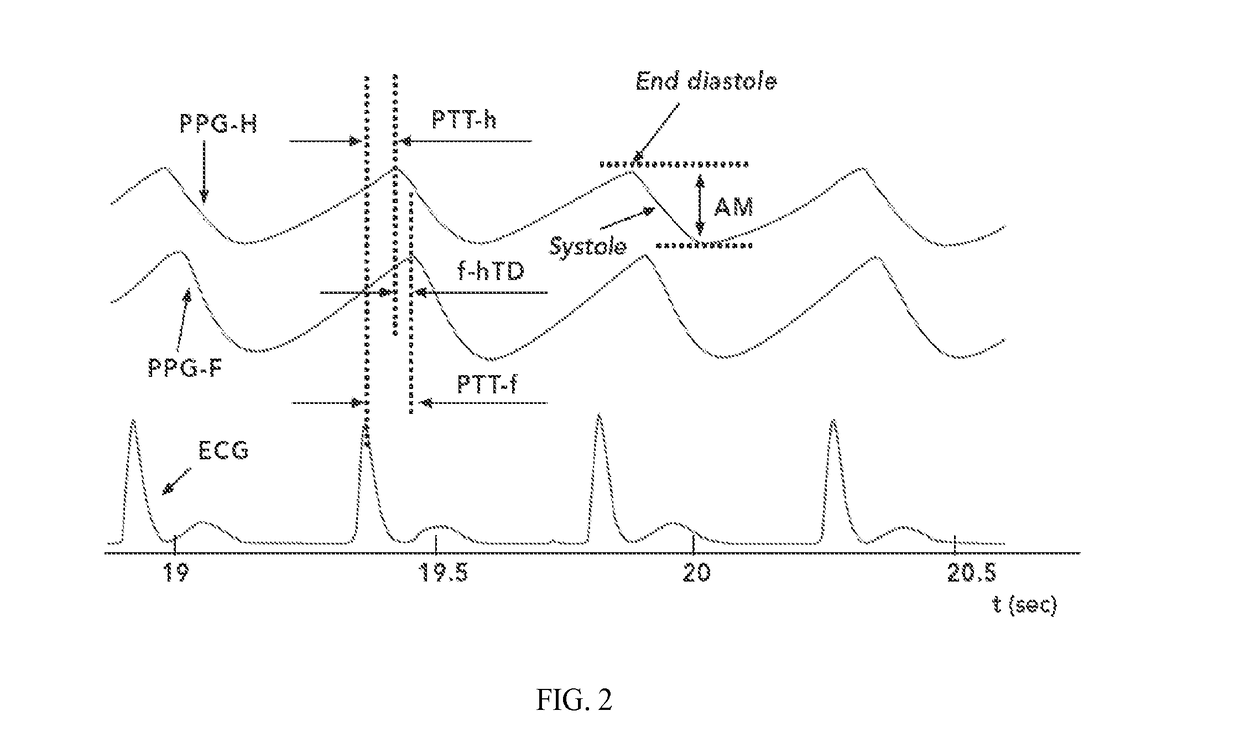
[0013]The present invention provides methods for detecting the likelihood of patent ductus arteriosus (PDA) in an infant comprising a) obtaining or receiving electrocardiogram (ECG) signals from the infant; b) obtaining or receiving photoplethysmographic (PPG) signals from a site on the upper body (UB) of the infant, and optionally from a foot (F) of the infant; c) determining, for each PPG pulse the PPG pulse amplitude (AM) between the end-diastolic maximum and the following minimum of systolic decrease for the UB PPG pulses; d) determining the mean of one or more of the following parameters for a plurality of the PPG pulses in the selected section: (i) a relative pulse amplitude (rAM) by dividing the AM by the systolic decrease minimum to obtain a rAM for the upper body (rAM-UB) PPG pulses; (ii) a pulse transit time (PTT-UB) between an R wave of the ECG and the onset of systolic decrease for the corresponding UB PPG pulse; (iii) a ratio PTT-UB/rAM-UB between the pulse transit time (PTT-UB) and the relative pulse amplitude for the UB (rAM-UB) PPG pulse; (iv) a ratio rAM-UB/PTT-UB between the relative pulse amplitude for the UB (rAM-UB) PPG pulse and the pulse transi

Problems solved by technology

Patent ductus arterious (PDA) is a common problem causing significant morbidity and mortality in preterm infants.
A PDA with a significant left to right shunting can lead to increased neonatal morbidity such as respiratory distress, cardiac failure, low blood pressure, and decreased peripheral organ perfusion, and leads to an increased incidence of intraventricular hemorrhage, necrotizing enterocolitis and chronic lung disease (Sehgal and McNamara 2012).
Assessment of the significance of the ductal flow is challenging.
The difficulty in precisely separating the pathological ductus arteriosus from the innocent ductus arteriosus may be due to the lack of scientific evidence of benefit or causality (McNamara and Sehgal 2007, Sehgal and McNamara 2012).
The ensuing effect is medical ambiguity and an unending dispute of whether these neonates have to be treated, the optimum timing of treatment and implications to when intervention is most effective.
The reasons for this apparent lack of effect may relate to a lack of relationship of a HSDA to neonatal outcomes due to inaccurate assignment of hemodynamic significance.
Other than clinical parameters and echocardiographic evaluation, there are no other non-invasive clinical tools to help assess this common but difficult diagnostic entity from a therapeutic standpoint.
Echocardiographic evaluation has been considered the gold standard for diagnosis of a patent ductus arteriosus, but quantifying the size of ductal flow which leads to negative consequences remains a problem (McNamara and Sehgal 2007, Evans et al.
The current definition of HSDA is not adequate since it is entirely reliant on size (McNamara and Sehgal 2007).
The lack of a uniform approach is a key hindrance towards better comprehension of the clinical effect of a patent ductus.
However, flow indicators hav

Method used


Image

  • Detection of patent ductus arteriosus using photoplethysmography
  • Detection of patent ductus arteriosus using photoplethysmography
  • Detection of patent ductus arteriosus using photoplethysmography

Examples


Embodiment Construction

[0023]The invention provides a method for detecting the likelihood of a patent ductus arteriosus (PDA) in an infant comprising:

[0024]a) obtaining or receiving electrocardiogram (ECG) signals from the infant;

[0025]b) obtaining or receiving photoplethysmographic (PPG) signals from a site on the upper body (UB) of the infant, and optionally from a foot (F) of the infant;

[0026]c) determining, for each PPG pulse the PPG pulse amplitude (AM) between the end-diastolic maximum and the following minimum of systolic decrease for the UB PPG pulses;

[0027]d) determining the mean of one or more of the following parameters for a plurality of the PPG pulses in the selected section:[0028](i) a relative pulse amplitude (rAM) by dividing the AM by the systolic decrease minimum to obtain a rAM for the upper body (rAM-UB) PPG pulses;[0029](ii) a pulse transit time (PTT-UB) between an R wave of the ECG and the onset of systolic decrease for the corresponding UB PPG pulse;[0030](iii) a ratio PTT-UB / rAM-UB...


PUM


Abstract

Methods and systems are described for detecting the likelihood of patent ductus arteriosus (PDA) in an infant using electrocardiogram and photoplethysmographic pulse signals obtained from the upper body and foot of the infant.

Description

CROSS-REFERENCE TO RELATED APPLICATIONS[0001]This application claims the benefit of U.S. Provisional Patent Application No. 62 / 042,823, filed on Aug. 28, 2014, and of U.S. Provisional Patent Application No. 62 / 134,619, filed on Mar. 18, 2015, the contents of which are incorporated herein by reference.BACKGROUND OF THE INVENTION[0002]Throughout this application various publications are referred to in parentheses. Full citations for these references may be found at the end of the specification. The disclosures of these publications are hereby incorporated by reference in their entirety into the subject application to more fully describe the art to which the subject invention pertains.[0003]Patent ductus arterious (PDA) is a common problem causing significant morbidity and mortality in preterm infants. The ductus arteriosus is a blood vessel that connects the aorta and pulmonary artery and plays an important role in fetal life. In full term newborn infants, the ductus arteriosus constr...

Claims


Application Information

Patent Timeline
IPC IPC(8): A61B5/024A61B5/00A61B5/0452A61B5/02A61B5/352A61B5/366
CPCA61B5/02416A61B5/02007A61B2503/045A61B5/0452A61B5/6801A61B5/6829A61B5/7282A61B5/6814A61B5/6825A61B2503/04A61B5/366A61B5/352A61B5/349
Inventor KOPPEL, ROBERT
Owner THE FEINSTEIN INST FOR MEDICAL RES
Why Eureka
Try Eureka