Central Pressurized Cadaver Model

a cadaver model and central pressurization technology, applied in the field of medical equipment, can solve the problems of affecting the application of virtual reality simulation in the training of resuscitative endovascular balloon occlusion of the aorta, requiring extensive training, and complicated execution of the aorta resuscitation operation

Active Publication Date: 2017-09-28
UNIV OF MARYLAND BALTIMORE
View PDF19 Cites 1 Cited by
  • Summary
  • Abstract
  • Description
  • Claims
  • Application Information

AI Technical Summary

Benefits of technology

[0012]The present invention is also directed to a training system for a surgical procedure that involves hemorrhagic shock. The training system comprises a cadaver and means for ligating veins of the cadaver. The training system also comprises a drainage tube disposed in an internal jugular vein of the cadaver and pressure tubing in a connecting relationship with the arterial catheter and an arterial catheter disposed in a brachial artery and a pressure transducer attached to the pressure tubing. One or more cannulas are disposed in a carotid artery, a common femoral artery or a combination thereof where one or more pressurized perfusion pumps are in a connecting relationship with the cannula. A blood replacement liquid is in a connecting relationship with the perfusion pumps.

Problems solved by technology

In a traumatic emergency, the execution of the resuscitative endovascular balloon occlusion of the aorta is highly complicated, thus requires extensive training.
However, the absence of percutaneous cannulation and / or open exposure of the groin, which are essential considerable components of resuscitative endovascular balloon occlusion of the aorta, hindered the application of virtual reality simulation in resuscitative endovascular balloon occlusion of the aorta trainings.
Animal testing provides dynamic blood flow and hemorrhage, but it lacks similarity to the access anatomy that is critical for mastering the resuscitative endovascular balloon occlusion of the aorta.
In addition, animal testing can be subjected to ethical concerns for reasons of animal welfares.
However, the most challenging aspect of using cadaver model for this training is the lack of pulsatile blood flow, which is essential to simulate traumatic injuries.
Particularly, the prior art is deficient in this aspect.

Method used


Image

  • Central Pressurized Cadaver Model
  • Central Pressurized Cadaver Model
  • Central Pressurized Cadaver Model

Examples


example 1

Preparation of the Cadaver Model

[0041]Fresh cadavers are refrigerated at 36° F. with no embalming performed. Arterial flushing is performed prior to refrigeration with boric acid solution (B4, Hydrol Chemical Co, Yeardon, Pa.) to keep the body at a pH of 7.4. A body that has no, or minimal history of, severe peripheral vascular disease or vascular reconstruction is selected.

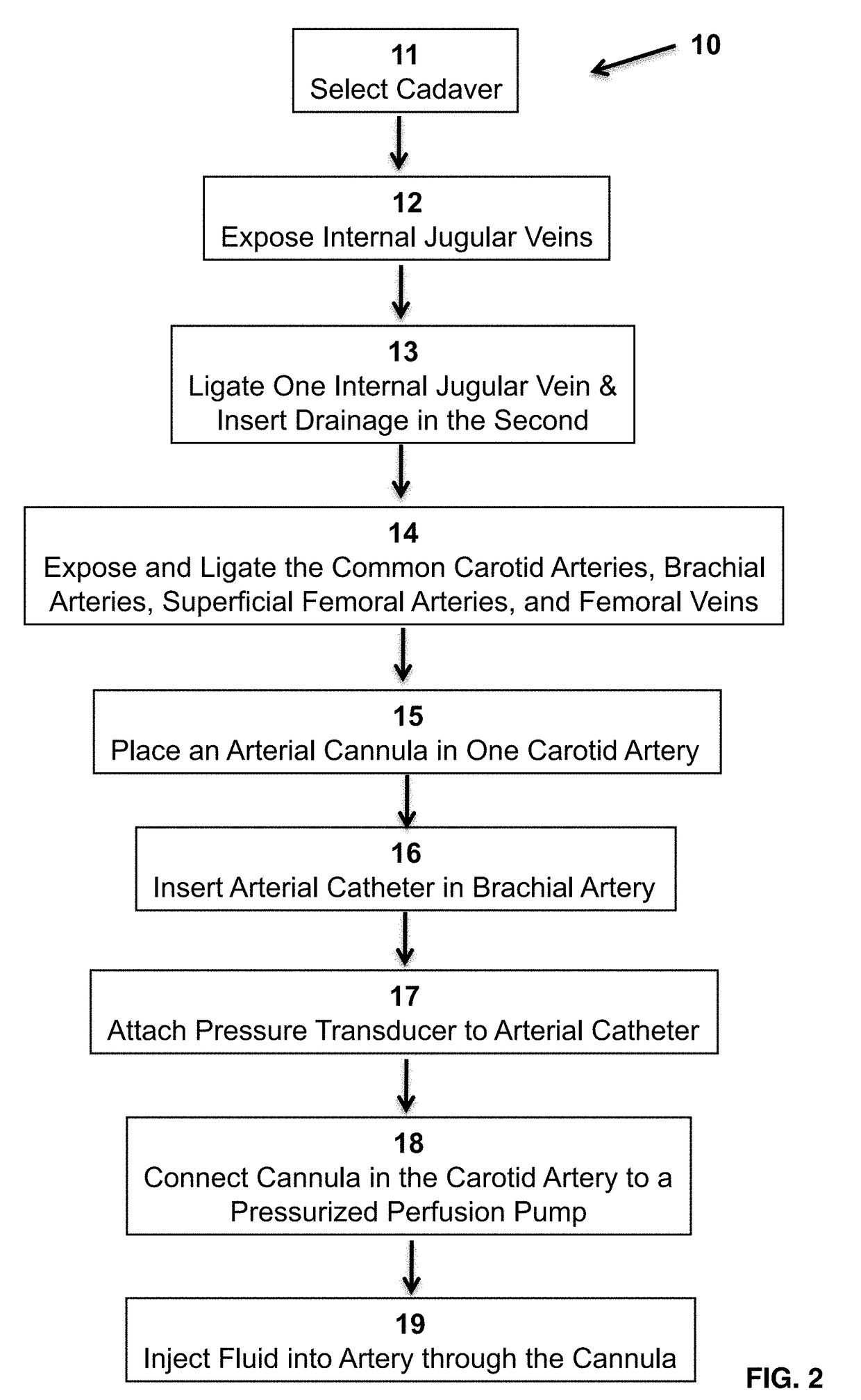
[0042]The selected cadaver is placed on a fluoroscopy table with the head at the sink to allow easy drainage of perfusion fluid, as shown in FIG. 3. The bilateral internal jugular veins, common carotid arteries, brachial arteries, superficial femoral arteries, and femoral veins are exposed through appropriate incisions. A drainage tube 21 for intermittent drainage is placed in the right internal jugular vein as show in FIG. 4. An arterial cannula 22 for perfusate is placed in the right carotid artery (FIG. 5). The left internal jugular vein and the carotid arteries are ligated, as are the bilateral superficial fe...

example 2

Training Procedure of Resuscitative Endovascular Balloon Occlusion of the Aorta

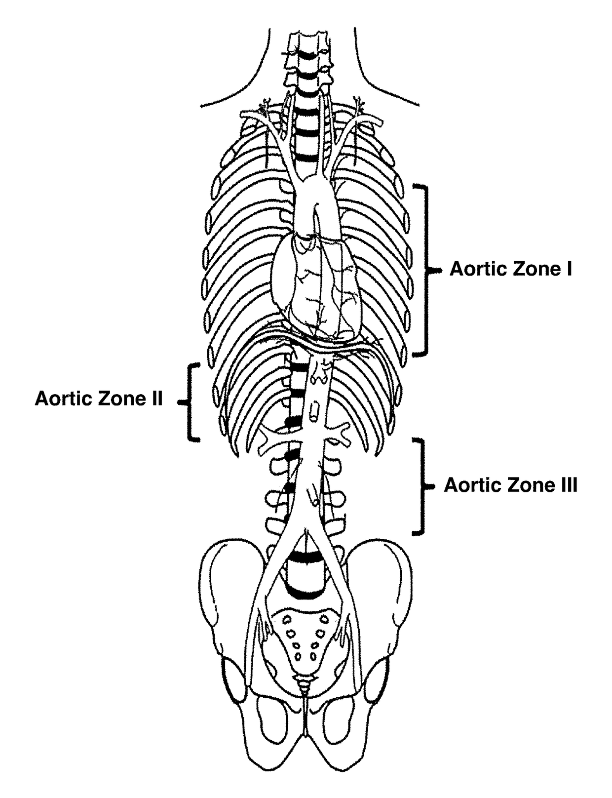
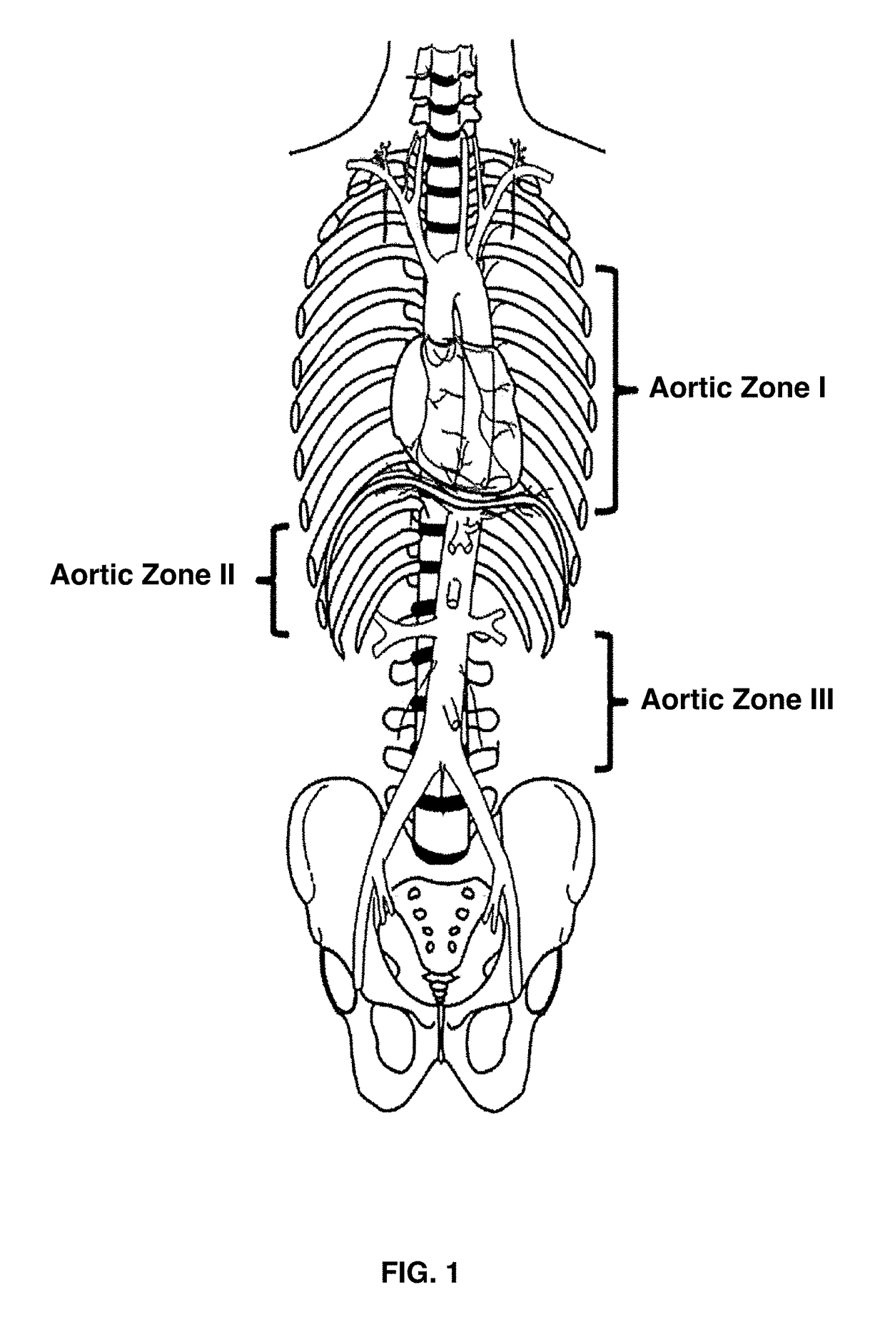
[0044]The Amplatz wire is advanced through the arterial catheter after approximation of insertion length by external landmarks. The arterial catheter is removed and a 12Fr sheath is inserted to allow access for the CODA balloon. The systolic blood pressure prior to inflation is about 40 mmHg to 80 mmHg, and infusate is pumped in a pulsatile fashion and titrated to this systolic blood pressure goal. If the systolic blood pressure rises about 80 mmHg, and infusate is pumped in a pulsatile fashion and titrated to his SBP goal. If the SBP rises above about 80 mmHg, prior to aortic occlusion, removal of fluid can be accomplished by venting the internal jugular cannula to allow drainage out of the central vasculature. A C-arm performs the function of a portable or digital X-ray machine by providing an image of the chest with wire 28 at the proximal descending thoracic aorta, as well as an image of the occlusive...


PUM


Abstract

Provided are methods for creating a pressurized cadaver model used for surgical procedure training. In the method the internal jugular veins, common carotid arteries, brachial arteries, superficial femoral arteries and femoral veins thereof of a cadaver are exposed. One internal jugular vein is ligated and a drainage tube is disposed in the other internal jugular vein. An arterial catheter is inserted into one brachial artery and connected to a pressure transducer and an arterial cannula is placed in a carotid artery and connected to a fusion pump. Fluid is injected into the artery through the cannulated carotid artery. A representative example of the surgical procedure includes resuscitative endovascular balloon occlusion of the aorta.

Description

CROSS-REFERENCE TO RELATED APPLICATION[0001]This international application claims benefit of priority under 35 U.S.C. 119(e) of provisional application U.S. Ser. No. 62 / 051,331, filed Sep. 17, 2014, the entirety of which is hereby incorporated by reference.BACKGROUND OF THE INVENTION[0002]Field of the invention[0003]The present invention generally relates to the field of medical equipment. More specifically, the invention relates to a central pressurized cadaver model for the Resuscitative Endovascular Balloon Occlusion of the Aorta procedure.[0004]Description of the Related Art[0005]The use of endovascular technology in the treatment of injury has steadily increased over the last several decades. Most recently, resuscitative endovascular balloon occlusion of the aorta (REBOA) has emerged as a potentially lifesaving technique for severely injured patients that do not respond to volume as a bridge to hemostasis. Many case series of resuscitative endovascular balloon occlusion of the ...

Claims


Application Information

Patent Timeline
Patent Type & Authority Applications(United States)
IPC IPC(8): G09B23/30
CPCG09B23/306G09B23/303
Inventor BRENNER, MEGANSCALEA, THOMAS
Owner UNIV OF MARYLAND BALTIMORE
Why Eureka
Try Eureka