
Medical device imagable by ultrasound

a medical device and ultrasound technology, applied in the field of medical devices, can solve the problems of not providing sufficient contrast for accurate imaging, present health concerns for patients and clinicians, etc., and achieve the effects of enhancing ultrasound echogenicity, good contrast, and effective monitoring

Inactive Publication Date: 2017-10-12
TYCO HEALTHCARE GRP LP
View PDF7 Cites 0 Cited by
  • Summary
  • Abstract
  • Description
  • Claims
  • Application Information

AI Technical Summary

Benefits of technology

The patent describes medical devices that can be easily seen using ultrasound. They contain material that releases gas to improve their image. The devices can be placed or moved through a patient's blood vessels and can be easily monitored by ultrasound. This eliminates the need for fluoroscopy and reduces the patient's exposure to radiation. The release of gas can also help the physician see the blood vessel in which the device is being used. Overall, this technology allows for better visualization of medical devices and structures using ultrasound.

Problems solved by technology

Fluoroscopy requires the use of radiation for visualization, which may present health concerns for both patients and clinicians.
However, many medical devices do not provide for sufficient contrast for accurate imaging via ultrasound.

Method used


Image

  • Medical device imagable by ultrasound
  • Medical device imagable by ultrasound
  • Medical device imagable by ultrasound

Examples


Embodiment Construction

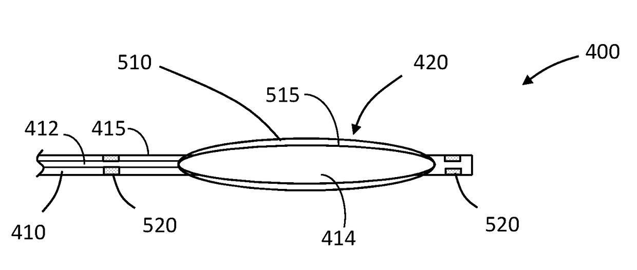
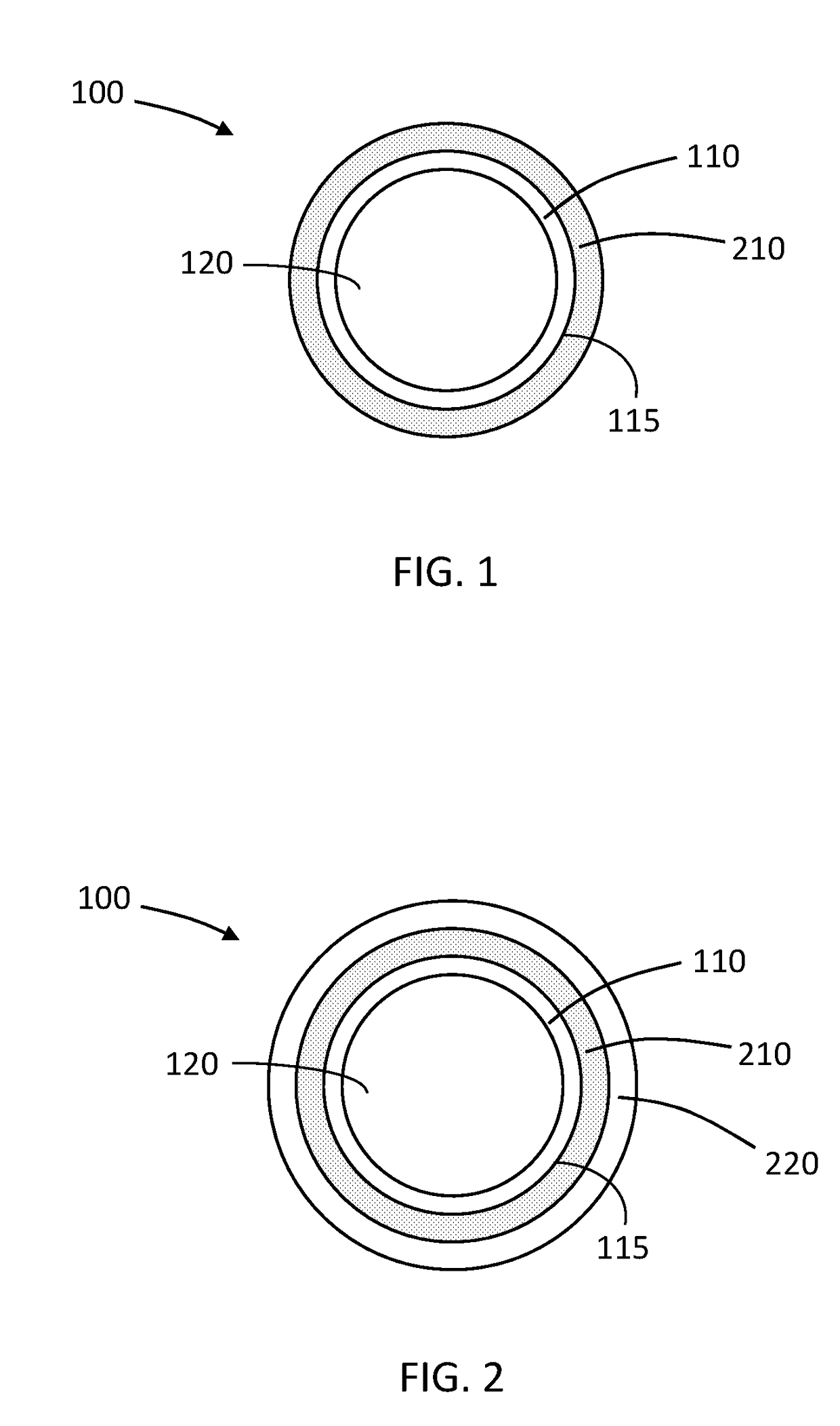
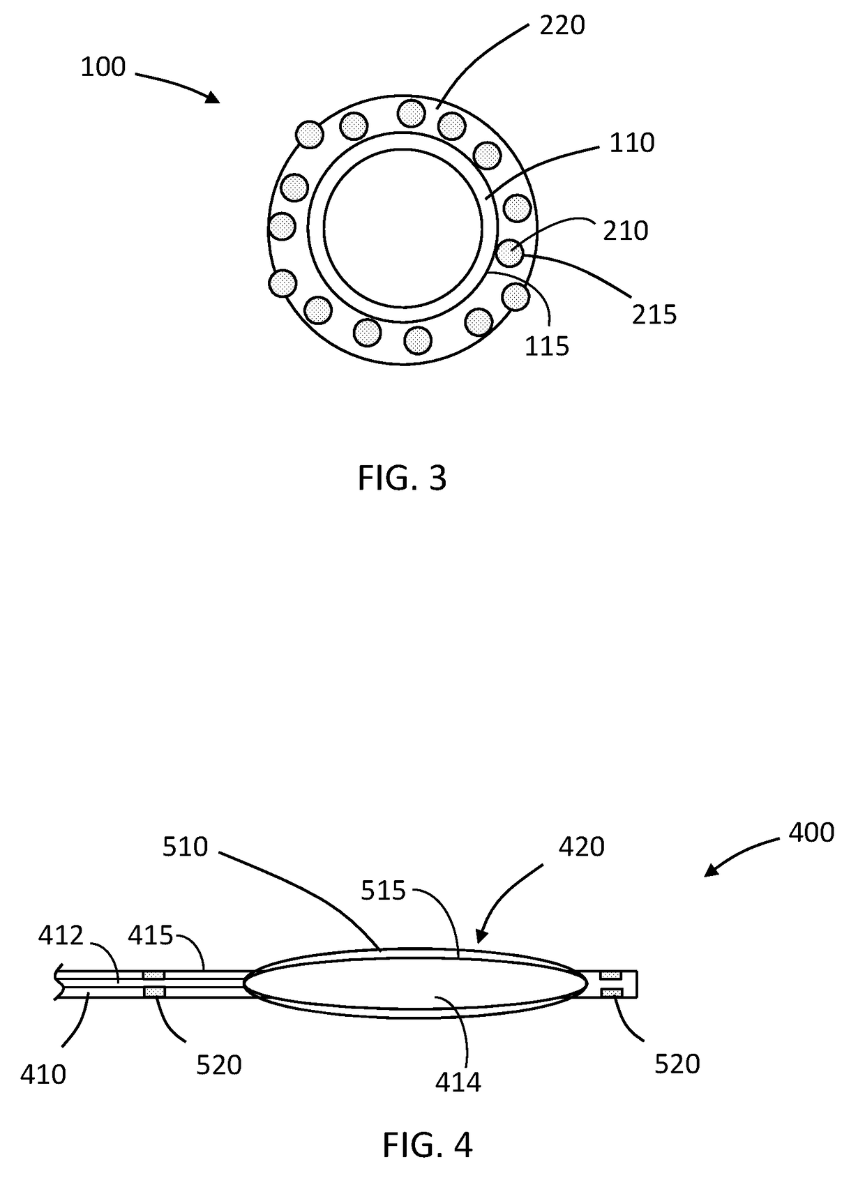
[0024]Reference will now be made in greater detail to various embodiments of the subject matter of the present disclosure, some embodiments of which are illustrated in the accompanying drawings. Like numbers used in the figures refer to like components, steps and the like. However, it will be understood that the use of a number to refer to a component in a given figure is not intended to limit the component in another figure labeled with the same number. In addition, the use of different numbers to refer to components in different figures is not intended to indicate that the different numbered components cannot be the same or similar to other numbered components.

[0025]The present disclosure describes, among other things, a medical device including a structural element comprising an outer surface and a material on the outer surface of the structural element. The material defines void spaces and contains a gas entrained within the void spaces. The material, upon contact with physiolog...


PUM

PropertyMeasurementUnit
solubilityaaaaaaaaaa
pressureaaaaaaaaaa
pressureaaaaaaaaaa

Abstract

A medical device includes a structural element comprising an outer surface and a material on the outer surface of the structural element. The material defines void spaces containing a gas entrained within the void spaces. The material, upon contact with physiological fluid, is configured to release the gas in an amount sufficient for the gas to be imaged by ultrasound.

Description

FIELD[0001]The present disclosure relates to, among other things, medical devices, or portions thereof, visible via ultrasound.TECHNICAL BACKGROUND[0002]Many medical procedures, whether interventional or diagnostic, include use of instruments that are, or have markers that are, visible by fluoroscopy. Fluoroscopy requires the use of radiation for visualization, which may present health concerns for both patients and clinicians.[0003]Ultrasound techniques, which do not employ radiation, provide an alternative to fluoroscopy. However, many medical devices do not provide for sufficient contrast for accurate imaging via ultrasound.BRIEF SUMMARY[0004]Described herein, among other things, are medical devices that are configured to provide good contrast for viewing by ultrasound. The devices include material that contains or releases gas to enhance ultrasound echogenicity. In some embodiments, the devices, or one or more portions thereof, are coated with a material that releases gas, such ...

Claims


Application Information

Patent Timeline
Patent Type & Authority Applications(United States)
IPC IPC(8): A61B8/08A61M25/10A61B90/00
CPCA61B8/0841A61B8/481A61B90/39A61M2202/0225A61B2090/3925A61M2025/1093A61M25/0108A61M25/10A61B8/12A61K49/223A61L29/085A61L29/18A61L2300/622A61L2400/10A61M2025/1075A61M2025/1088A61M2025/1031A61L29/145A61L29/146A61L29/148
Inventor MCPEAK, THOMAS
Owner TYCO HEALTHCARE GRP LP
Learn More