Tool for percutaneous joint cartilage destruction and preparation for joint fusion

a percutaneous joint and cartilage technology, applied in the field of medical devices, can solve the problems of relatively cumbersome methods and time-consuming, and achieve the effects of less time-consuming, less cumbersome, and softening the cartilage in the join

Active Publication Date: 2017-11-16
EXSOMED CORP
View PDF4 Cites 13 Cited by
  • Summary
  • Abstract
  • Description
  • Claims
  • Application Information

AI Technical Summary

Benefits of technology

[0006]A tool for destroying or removing cartilage from bone includes a proximal end, a distal end, and a cannula extending through the tool. The distal end includes at least one scraping and / or cutting surface to break up and / or soften the cartilage in the joint as the scraping and / or cutting surface is rotated or otherwise moved within the cavity. The proximal end can include a section that can be received by a device, such as a drill, that can be used to cause the tool to rotate. The cannula can be configured to receive a guide wire, such as a K-wire, to help guide the distal end of the tool towards and into the joint cavity to be scrubbed or arthrolysed.
[0007]When used, the tool is preferably attached to a drill and the cannula has received an end of a guide wire, wherein a portion of the guide wire has been inserted into the joint cavity. The drill rotates the tool around the guide wire and can be used to push the tool over the guide wire and into the joint cavity. Once the tool is inserted into the joint cavity, the drill can be used to move the tool along an axis of the guide wire into the cavity as well as cause the tool to rotate within the cavity.
[0008]Procedures using a tool as described herein can be less cumbersome and less time consuming, less traumatic compared to traditional medullary cavity scrubbing techniques. The device as designed preferably provides a lead cutting surface suitable for trans-cutaneous or percutaneous insertion into a joint though a small skin hole
[0009]The device being of a cannulated design allows for controlled delivery of the sharp cutting lead end of the device into and out of the joint, thus providing for cartilage destruction, but preventing creep of the device into other areas such as nerves and tendons

Problems solved by technology

Although this technique works relatively well, the method is relatively cumbersome and time consuming, requiring a large exposure or access to the joint.

Method used


Image

  • Tool for percutaneous joint cartilage destruction and preparation for joint fusion
  • Tool for percutaneous joint cartilage destruction and preparation for joint fusion
  • Tool for percutaneous joint cartilage destruction and preparation for joint fusion

Examples


Embodiment Construction

[0016]The description of exemplary embodiments of the present invention provided below is merely exemplary and is intended for purposes of illustration only; the following description is not intended to limit the scope of the invention disclosed herein. Moreover, recitation of multiple embodiments having stated features is not intended to exclude other embodiments having additional features or other embodiments incorporating different combinations of the stated features.

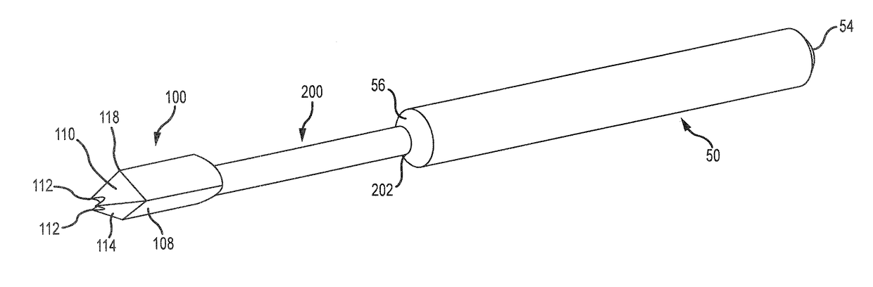
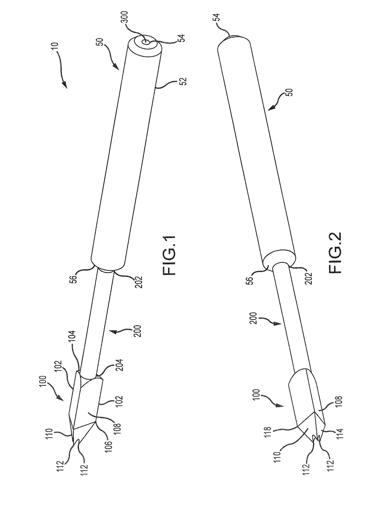
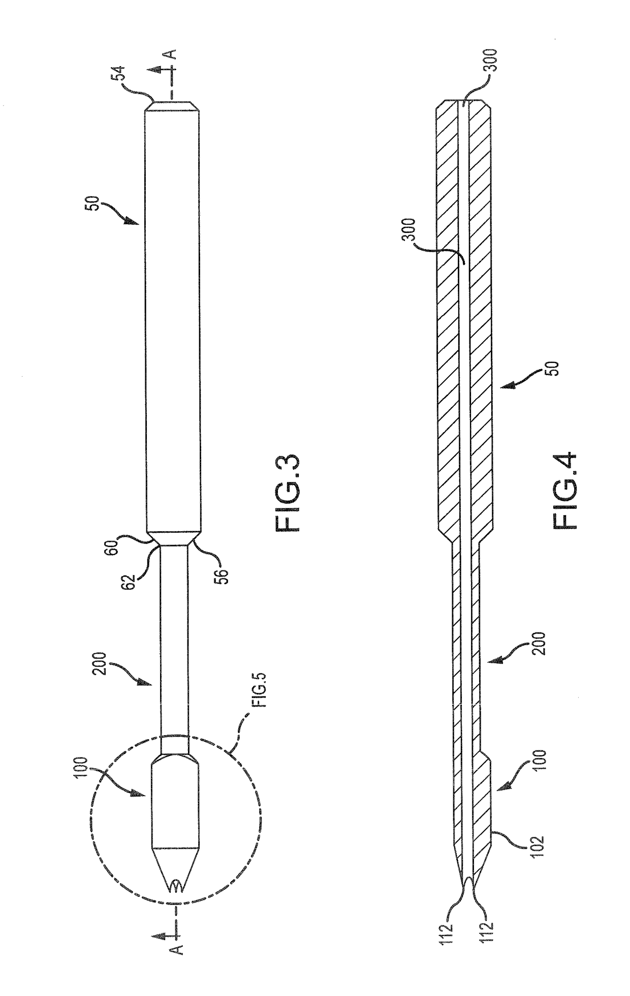
[0017]The present disclosure describes medical tools and systems that are suitable for use in treatment of joints, and particularly for treatments including joint fusion or arthrodesis, such as arthrodesis of phalanges. For example, exemplary devices and systems described herein are suitable for treatment of a distal interphalangeal (DIP) joint, and other joints such as for fusion and the scrubbing of the joint prior to implantation of another device, such as a bone screw. The devices and systems are conveniently des...


PUM


Abstract

A tool for scrubbing, removing tissue from the medullary cavity or joint cavity of a bone or removing cartilage and / or cortical bone from a joint cavity has a proximal end, a distal end, and a cannula extending therethrough. The distal end includes at least one scraping surface and / or cutting surface to soften marrow in the medullary cavity or joint cavity as the scraping and / or cutting surface(s) are rotated or otherwise moved relative to the medullary cavity or joint cavity. When used, the tool may be attached to a drill and the cannula receives a K-wire previously inserted into the medullary or joint cavity. The drill rotates the tool around the K-wire and pushes the tool over the K-wire into the medullary cavity or joint cavity.

Description

FIELD OF THE INVENTION[0001]The present disclosure generally relates to medical devices. More particularly, the disclosure relates to tools suitable for preparing a joint for fusion with a percutaneous method of cartilage destruction.BACKGROUND OF THE INVENTION[0002]For various reasons, it may be desirable to destroy or remove cartilage from the joint area of a bone. For example, such procedures are often employed in the treatment of hand or foot injuries during the process of joint fusion to facilitate the healing of a joint fusion. Joint fusion is indicated for unstable or painful joints. The process of joint fusion involves removal or destruction of the smooth cartilage lining of a joint followed by fixation of the raw ends of the bone to each other with the intent to have the bone ends heal and form a solid, non-movable, bone construct that is no longer unstable or painful.[0003]A typical method of performing joint cartilage removal involves creating and incision or open arthrot...

Claims


Application Information

Patent Timeline
Patent Type & Authority Applications(United States)
IPC IPC(8): A61B17/16
CPCA61B17/1615A61B17/164A61B17/1659A61B17/16A61B17/1604A61B17/1637A61B17/1662A61B17/1682
Inventor CHAMPAGNE, LLOYD P.ZOLDOS, JOZEFHUNSON, ATILLA A.
Owner EXSOMED CORP
Why Eureka
Try Eureka