
A medical placement alarm

a technology for medical devices and alarms, applied in the field of apparatus for providing medical devices for placement, can solve the problems of malpositioning or disruption of medical devices, increasing the risk of future device failures or complications, etc., and achieve the effect of improving techniqu

Active Publication Date: 2018-08-02
KONINKLJIJKE PHILIPS NV
View PDF3 Cites 1 Cited by
  • Summary
  • Abstract
  • Description
  • Claims
  • Application Information

AI Technical Summary

Benefits of technology

The patent is about a device and method that can help doctors check the position of a second medical device during implantation. This can help reduce the risk of the second device getting caught or trapped under the first device. This can be done before or during the implant procedure.

Problems solved by technology

Mistakes made in this procedure can lead to malpositioning or disruption of the medical device following the implantation.
This increases the risk of future device failures or complications.

Method used


Image

  • A medical placement alarm
  • A medical placement alarm
  • A medical placement alarm

Examples


Embodiment Construction

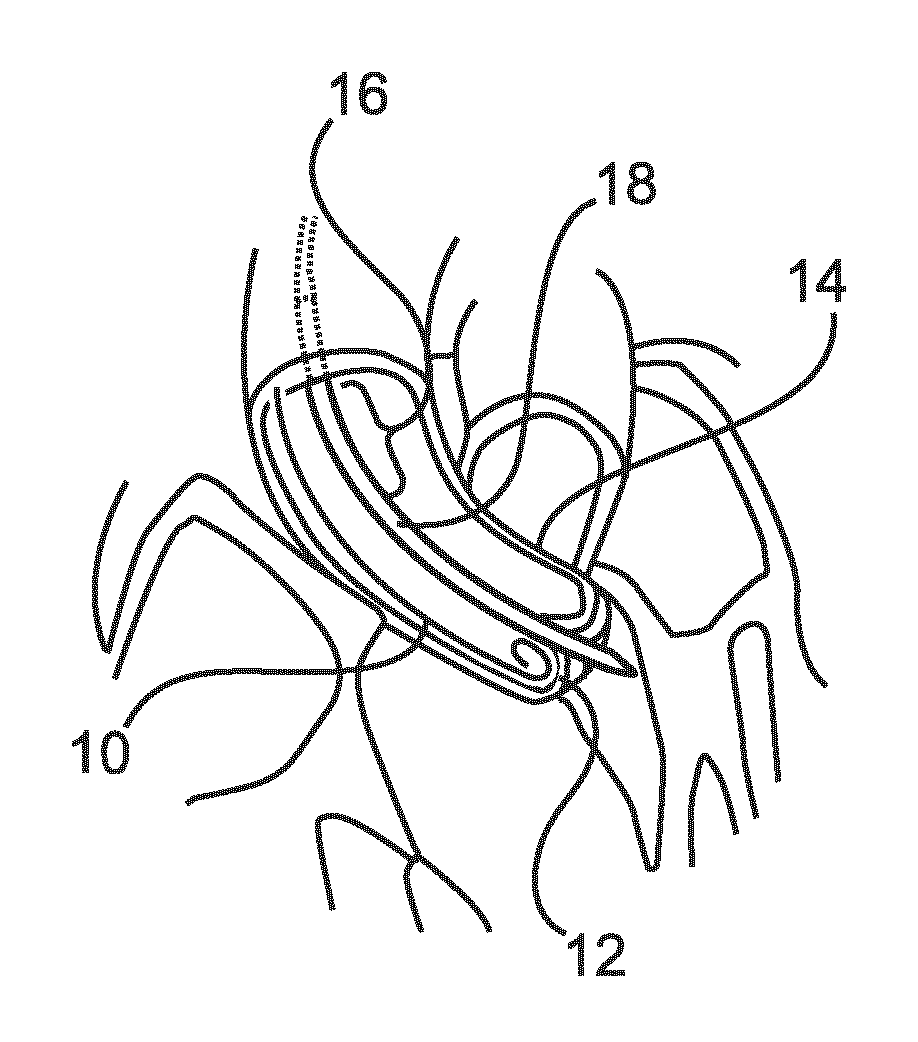
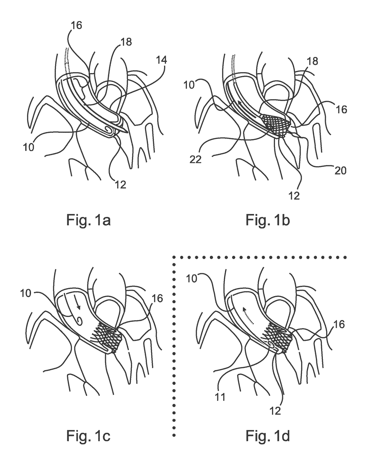
[0034]The placement of a medical device, for example a valve prosthesis during a TAVI procedure, is highly dependent on accurate positioning of the valve prosthesis on the supporting anatomy, for a successful outcome.

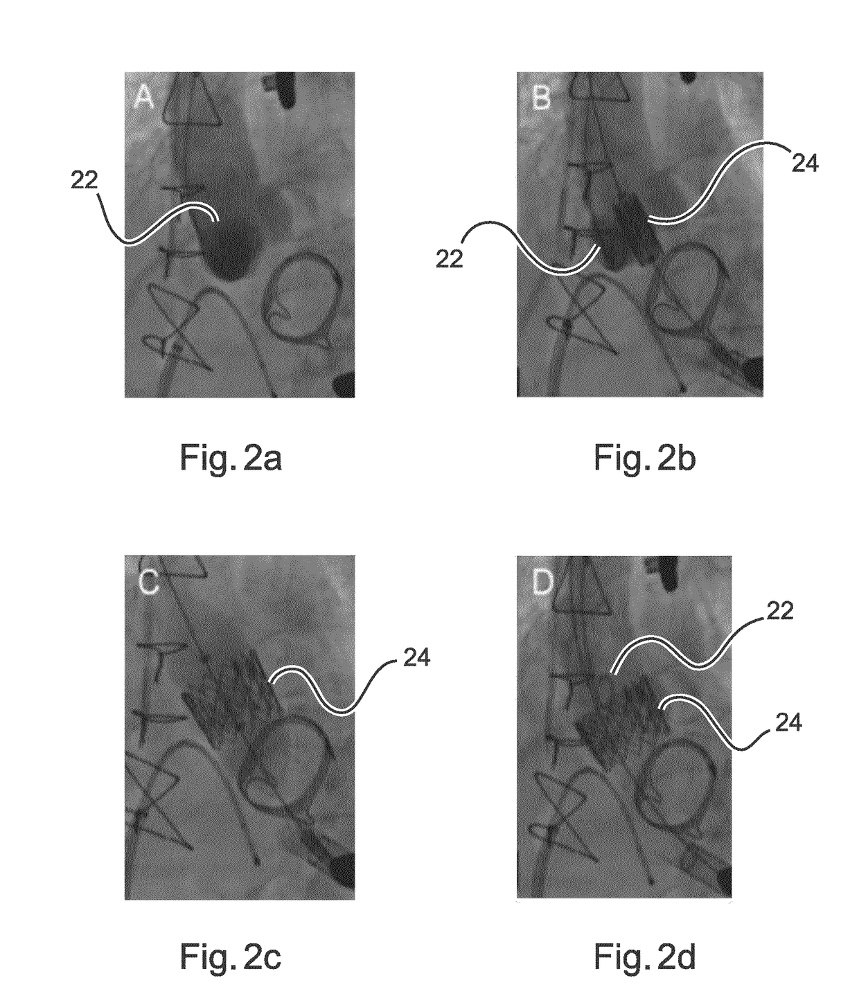
[0035]Classically, once a valve prosthesis has been placed in an implantation position, and the position of the valve has been confirmed using an angiogram, a classic valve deployment consists of the following steps:

[0036]Firstly, a heart pacemaker attached to the heart is put into a so-called “hyperpacing” mode to reduce the magnitude of the patient's heartbeat. Secondly, the valve prosthesis is deployed. In some types of valve, for example the Medtronic CoreValve™ the interventionist releases the proximal part of the prosthesis, confirms the position, pulls a pigtail catheter back, and then proceeds until the valve is fully deployed. In another type of valve deployment, for example the Edwards SAPIEN XT™ valve, the interventionist withdraws the pigtail catheter, and t...


PUM


Abstract

Many medical procedures involve the implantation of an element in a vascular body. Current imaging techniques allow the positioning of such expandable elements to be checked, before and after implantation, using contrast bursts into the blood from a pigtail catheter. When the implantation of the medical device needs to be performed quickly, the pigtail catheter is pulled back briskly. This process is prone to error, because at the point of implantation, a medical professional has many other factors to consider, as well as the withdrawal of the catheter. If an implantable device traps the pigtail catheter during or after expansion, undesirable results can occur, such as having to pull sharply on the catheter to remove it, and in the process, disrupting the newly implanted device. This application discusses a technique for automatically generating a region of implantation around a medical device prior to and during implantation. If a second medical device, such as a pigtail catheter, strays into the area, an alarm can be generated to warn a medical professional to withdraw the catheter, before deployment continues.

Description

FIELD OF THE INVENTION[0001]The present invention relates to an apparatus for providing a medical placement alarm, a method for providing a medical placement alarm, an X-ray imaging arrangement, a computer program element, and a computer-readable medium.BACKGROUND OF THE INVENTION[0002]A medical device implantation procedure is used when it is necessary to implant a medical device inside a patient. Typically, a guide wire or intervention device, supporting a medical device to be implanted, is arranged in a lumen of a patient. A catheter can be used to emit bursts of contrast medium, detectable using a suitable fluoroscopic approach, to better visualize the anatomy. During such an intervention, an implant is deployed very quickly. The point of implantation or deployment of the medical device is a critical time period. Many actions need to be performed by a medical professional, in quick succession. Mistakes made in this procedure can lead to malpositioning or disruption of the medica...

Claims


Application Information

Patent Timeline
IPC IPC(8): A61B6/12A61B6/00
CPCA61B6/12A61B6/487A61B6/481G06T7/215A61B2034/2065G06T2207/10121A61B90/39A61B2090/376G06T2207/30101A61B6/102
Inventor PIZAINE, GUILLAUME JULIEN JOSEPHNEMPONT, OLIVIER PIERREAUVRAY, VINCENT MAURICE ANDREFLORENT, RAOUL
Owner KONINKLJIJKE PHILIPS NV
Learn More