Method for displaying an anatomical image of a coronary artery on a graphical user interface

an anatomical image and user interface technology, applied in the field of anatomical images of blood vessels, can solve the problems of difficult identification of the cross-section of the ivus or oct image, difficult to guide the catheter to the lesion using only ivus or oct, etc., to improve the understanding improve the accuracy of the anatomical image, and improve the effect of user experien

Active Publication Date: 2018-09-27
CANON USA
View PDF15 Cites 10 Cited by
  • Summary
  • Abstract
  • Description
  • Claims
  • Application Information

AI Technical Summary

Benefits of technology

[0007]The present disclosure is directed to a method for displaying an anatomical image to enhance a user's understanding of the anatomical image of a blood vessel to quickly interpret structural and molecular information obtained from an intravascular imaging system. The user is able to interpret the information obtained from an intravascular imaging system without displaying or reviewing...

Problems solved by technology

However, one reason why IVUS and OCT are not commonly used in a PCI procedure is that it is difficult to guide a catheter to a lesion using only IVUS or OCT.
Furthermore, when viewing the intravascular image frame of a coronary artery, it may be difficult for an interventional cardiologist to determine what location along the longitudinal silhouette of the coronary artery in an angiography image the IVUS or OCT image was acquired.
In other words, the cross-section of the IVUS or OCT image is difficult to locate when viewing an anatomical image of t...

Method used


Image

  • Method for displaying an anatomical image of a coronary artery on a graphical user interface
  • Method for displaying an anatomical image of a coronary artery on a graphical user interface
  • Method for displaying an anatomical image of a coronary artery on a graphical user interface

Examples


Embodiment Construction

[0037]The following description is of certain illustrative embodiments, although other embodiments may include alternatives, equivalents, and modifications. Additionally, the illustrative embodiments may include several novel features, and a particular feature may not be essential to practice the devices, systems, and methods described herein.

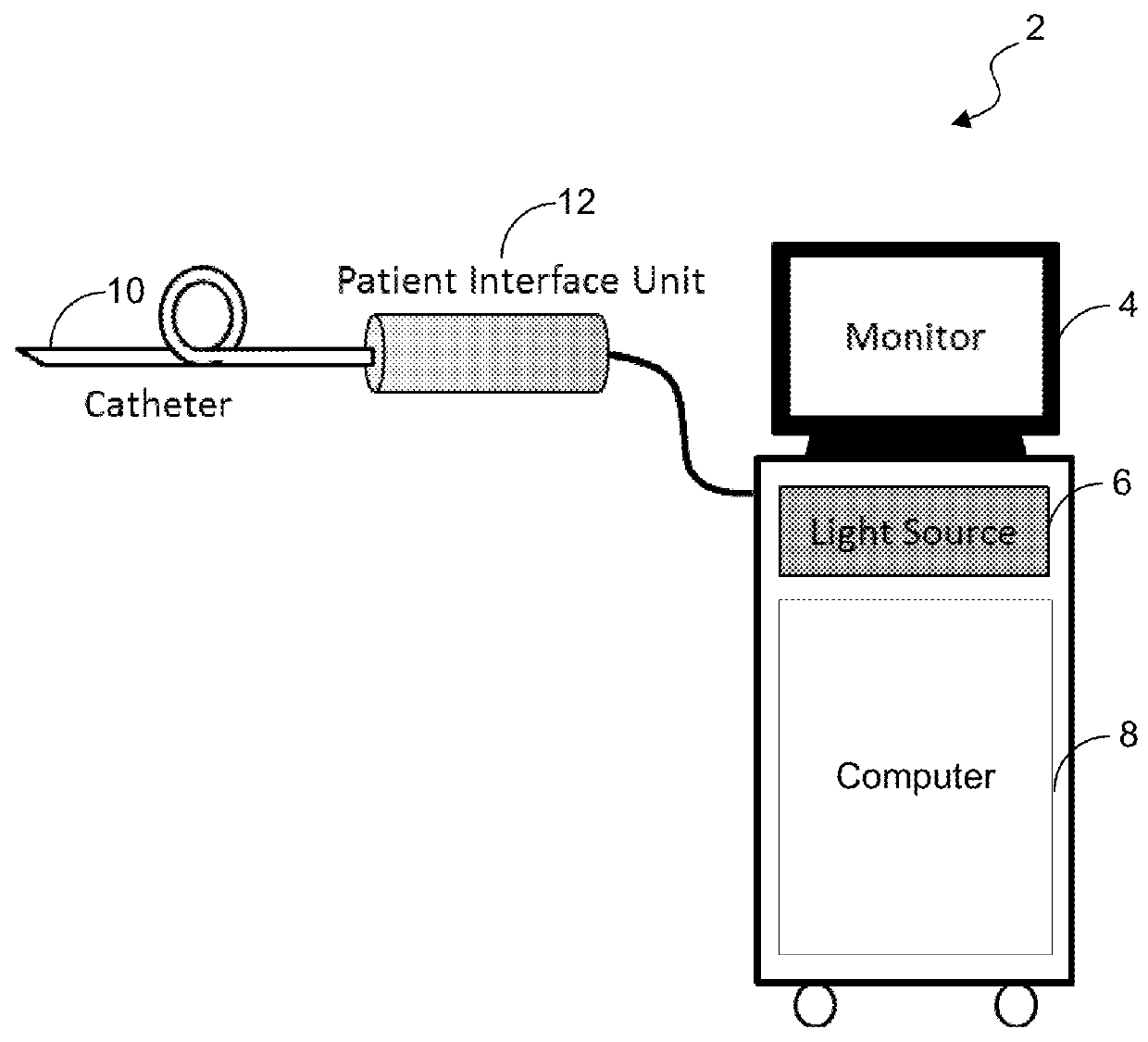
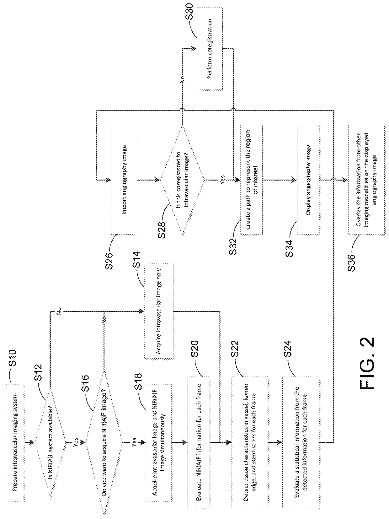
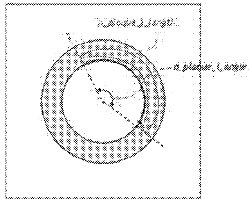
[0038]The present disclosure includes a feature for displaying an anatomical image to assist a user during review of the anatomical image of a blood vessel to quickly interpret structural and molecular information obtained from an intravascular imaging system. The user may interpret or review the information obtained from an intravascular imaging system without having to review an intravascular image frame. The present disclosure is directed to overlaying qualitative and quantitative information obtained from the intravascular imaging system onto the anatomical image of the blood vessel in a manner in which a user may more efficiently interpret...


PUM


Abstract

A method for displaying an anatomical image of a coronary artery on a graphical user interface with information acquired from a plurality of intravascular image frames. The method may include detecting qualitative information from the plurality of intravascular image frames, creating one or more indicator(s) from the qualitative information detected, and determining a spatial relationship between the anatomical image and a plurality of acquisition locations of the plurality of intravascular image frames and generating its linear representation. The method also includes displaying the anatomical image of the coronary artery with the linear representation overlaid thereon on a display device and overlaying the one or more indicator(s) representing at least one type of qualitative information on the anatomical image along the linear representation.

Description

CROSS-REFERENCE TO RELATED APPLICATION(S)[0001]This application relates, and claims priority, to U.S. Provisional Application Ser. No. 62 / 474,248, filed Mar. 21, 2017, the entire disclosure of which is incorporated by reference herein in its entirety.BACKGROUNDField of the Disclosure[0002]The present disclosure relates in general to an anatomical image of a blood vessel enhanced with structural and molecular information, and more particularly, to a method for displaying an anatomical image of a coronary artery with qualitative and quantitative information from an intravascular imaging system overlaid on the anatomical image.Description of the Related Art[0003]Percutaneous coronary intervention (PCI) has been improved by innovative imaging modalities including coronary angiography and intravascular imaging. Coronary angiography is a medical imaging technique used to visualize lumen of blood vessels and organs of the body. Coronary angiography is also used to guide a catheter insertio...

Claims


Application Information

Patent Timeline
IPC IPC(8): A61B90/00A61B17/3207A61B6/12A61B34/20A61B5/00A61B6/00
CPCA61B90/37A61B17/32075A61B6/12A61B34/20A61B5/0066A61B5/0084A61B6/481A61B6/504A61B2090/364A61B2034/2065A61B2090/3735A61B2090/376A61B2090/372A61B2017/00199A61B2017/00292A61F2/82A61B5/743
Inventor KUNIO, MIE
Owner CANON USA
Why Eureka
Try Eureka