Device, system and method for skin detection

a technology of skin detection and remote photoplethysmography, applied in the field of remote photoplethysmographic systems and devices, can solve the problems of not being robust to changes in ambient light color, not being able to detect skin areas under low illumination conditions or in darkness, and achieve reliable, accurate and fast skin detection.

Active Publication Date: 2019-02-21
KONINKLJIJKE PHILIPS NV
View PDF0 Cites 8 Cited by
  • Summary
  • Abstract
  • Description
  • Claims
  • Application Information

AI Technical Summary

Benefits of technology

[0015]It is an object of the present invention to provide a device and a corresponding method as well as a system which allow a reliable, accurate and fast detection of skin, in particular for use in a device and method for detecting vital signs of a subject.
[0028]The present invention is based on the idea to distinguish a pulse signal from noise in the frequency domain, for instance by evaluating the distribution of the energy in a sorted frequency domain signal derived from image data of a scene potentially including a skin are of a living being or even a complete living being or different body parts showing skin areas and non-skin areas. It has particularly been found that the energy of a pulse signal (in a sorted (or ranked) frequency domain representation) drops faster than the energy of the sorted (or ranked) noise spectrum. With a classifier, that is preferably trained accordingly e.g. using such ranked spectra as input, a reliable decision can be made whether or not a particular region of interest (ROI) in the image data, e.g. an image segment of the image frames of said image data, shows skin or not.
[0034]In another embodiment said sorting unit is configured to divide said spectral signal into two or more spectral sub-signals and to separately sort said sub-signals to obtain sorted spectral sub-signals representing the descriptor. It has been found that such sub-signals even better allow distinguishing e.g. a pulse signal from noise and, thus, skin from non-skin.
[0038]It has been found that a repetition of the transformation and sorting transforms a (peaked) pulse spectrum into a flat spectrum, while a (relatively flat) noise spectrum is transformed into a peaked spectrum. In the repeated transforms the phase information, the location of the peak, and the amplitude of the spectrum are preferably disregarded. The sorting makes sure that the peak is substantially at the same location improving the classification.
[0040]Further, in an embodiment said extraction unit is configured to combine, in particular to average, image data values of a group of pixels of said image data per image frame to obtain said PPG signal from said combined image data values. Hence, the average (combination) over a group of pixels may be taken and the time-evolution of concatenated averages may be considered as the 1D time signal. This improves the reliability of the classification. Basically, a loss of resolution (of the skin-map) is treated for an improved reliability of the classification because the averaging increases the difference between pulse and noise (by making the pulse signal less noisy).

Problems solved by technology

Therefore, remote photoplethysmographic systems and devices are considered unobtrusive and well suited for medical as well as non-medical everyday applications.
However, they are not robust to changes of ambient light color, which will change the color of light reflected from a skin area, and are not able to detect skin areas under low illumination conditions or in darkness.
Moreover, such methods cannot always differentiate skin from other surfaces with the same color.
However, the reliability of the approach depends on the robustness of PPG signal extractions, thus it is impacted by motion of a subject and the blood perfusion level.
Therefore, if a pulse signal is not periodic or weak, a camera-based system will have difficulties to detect / segment the skin areas.
Moreover, the approach is computationally expensive.
However, particularly for applications with low light level or in the dark, e.g. for night-time patient monitoring, the relatively low amplitude of these absorption variations, particularly in the near-infrared (NIR) wavelength range, is still problematic and causes practical attempts to fail distinguishing skin and non-skin.

Method used


Image

  • Device, system and method for skin detection
  • Device, system and method for skin detection
  • Device, system and method for skin detection

Examples


first embodiment

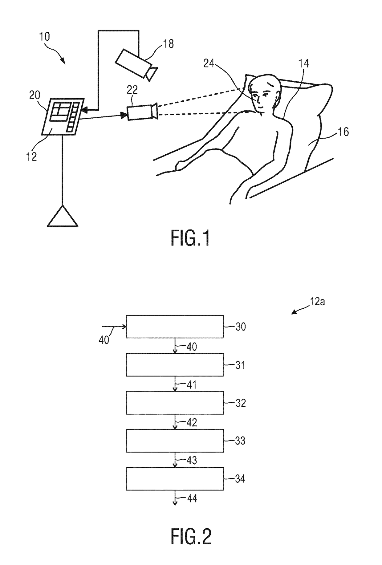
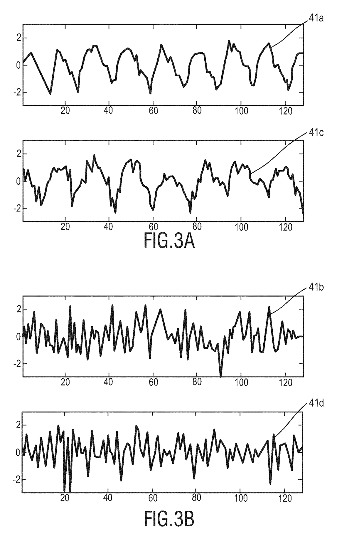
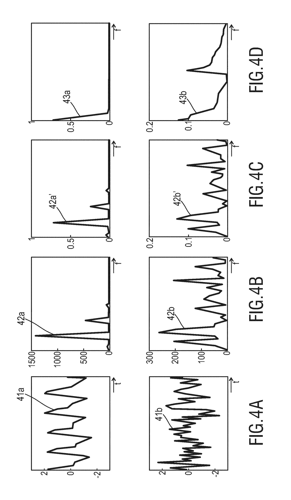
[0062]FIG. 2 shows a schematic diagram of a device 12a according to the present invention, which may be used as device 12 in the system 10 shown in FIG. 1. For deriving one or more vital signs of the subject 14 a skin area of the subject has to be found in the image data. For this purpose, the proposed device 12a comprises an input interface 30 for obtaining image data 40 of a scene, said image data comprising a time sequence of image frames acquired by the imaging unit 18. An extraction unit 31 extracts a PPG signal 41 from a region of interest of said image data, wherein said region of interest may a single pixel or a group of pixel or an area resulting from a segmentation of one or more image frames. A transformation unit 32 transforms said PPG signal 41 into a spectral signal 42. A sorting unit 33 sorts said spectral signal 42 to obtain a sorted spectral signal 43 representing a descriptor. Finally, a classifier 34 classifies said region of interest as skin region of a living be...

second embodiment

[0073]FIG. 5 shows a schematic diagram of a device 12b according to the present invention. In this embodiment a control unit 35 is provided for controlling said transformation unit 32 and said sorting unit 33 to perform two or more iterations, wherein the sorted spectral signal 43 output from said sorting unit 33 is used as input PPG signal 41′ for the transformation unit 32 in the next iteration. Thus, according to this embodiment a multiscale iteration may be performed as will be explained in the following.

[0074]With the first embodiment of the device 12a a transformed signal SL / 2 is obtained given the input PPG signal {right arrow over (P)}L, where pulse and noise have self-unified but mutually different interpretations. If the descriptor for pulse and noise is compared, the pulse-descriptor has a salient feature (e.g., peak at first location), whereas the noise does not. To obtain better classification performance, the descriptors from different classes require large between-cla...


PUM


Abstract

The present invention relates to a device, system and method for skin detection. To enable a reliable, accurate and fast detection the proposed device comprises an input interface (30) for obtaining image data of a scene, said image data comprising a time sequence of image frames, an extraction unit (31) for extracting a photoplethysmography (PPG) signal from a region of interest of said image data, a transformation unit (32) for transforming said PPG signal into a spectral signal, a sorting unit (33) for sorting said spectral signal to obtain a sorted spectral signal representing a descriptor, and a classifier (34) for classifying said region of interest as skin region of a living being or as non-skin region based on the descriptor.

Description

FIELD OF THE INVENTION[0001]The present invention relates to a device, system and method for skin detection.BACKGROUND OF THE INVENTION[0002]Vitas signs of a person, for example the heart rate (HR), the respiration rate (RR) or the arterial blood oxygen saturation (SpO2), serve as indicators of the current health state of a person and as powerful predictors of serious medical events. For this reason, vital signs are extensively monitored in inpatient and outpatient care settings, at home or in further health, leisure and fitness settings.[0003]One way of measuring vital signs is plethysmography. Plethysmography generally refers to the measurement of volume changes of an organ or a body part and in particular to the detection of volume changes due to a cardio-vascular pulse wave traveling through the body of a subject with every heartbeat.[0004]Photoplethysmography (PPG) is an optical measurement technique that evaluates a time-variant change of light reflectance or transmission of a...

Claims


Application Information

Patent Timeline
Patent Type & Authority Applications(United States)
IPC IPC(8): G06T7/00G06K9/62A61B5/024A61B5/0295A61B5/026A61B5/00
CPCG06T7/0012G06K9/6284A61B5/02433A61B5/0295A61B5/0261G06K9/6298A61B5/7267A61B5/7203G06T2207/30088G06T2207/30076G06T2207/20216A61B5/0075A61B5/0077A61B5/02416A61B5/02438G06F18/10G06F18/2433
Inventor WANG, WENJINDE HAAN, GERARD
Owner KONINKLJIJKE PHILIPS NV
Why Eureka
Try Eureka