

Method and an apparatus for determining haemodynamic status
a technology of haemodynamic status and apparatus, which is applied in the field of methods and apparatus for determining haemodynamic status, can solve the problems of abnormal heart rhythm, rapid heartbeat, and insufficient information obtained from cardiac electrical signals to determine individual conditions, etc., and achieve the effect of maximising the available data
- Summary
- Abstract
- Description
- Claims
- Application Information
AI Technical Summary
Benefits of technology
Problems solved by technology
Method used
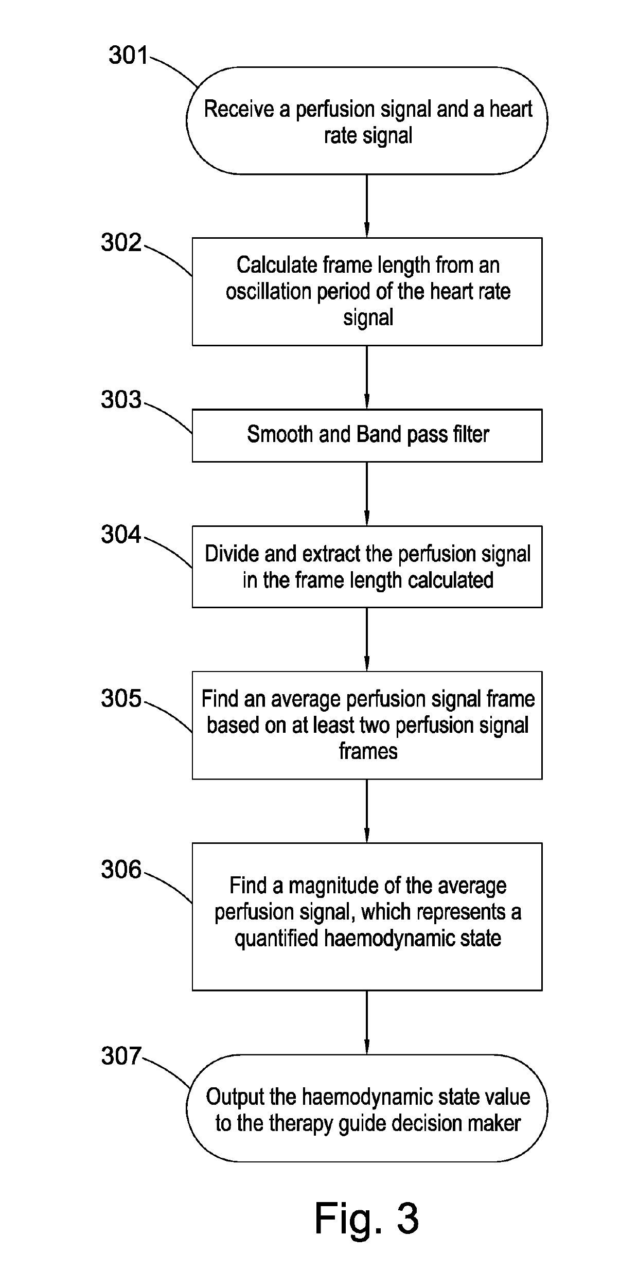
Image


Examples
Embodiment Construction
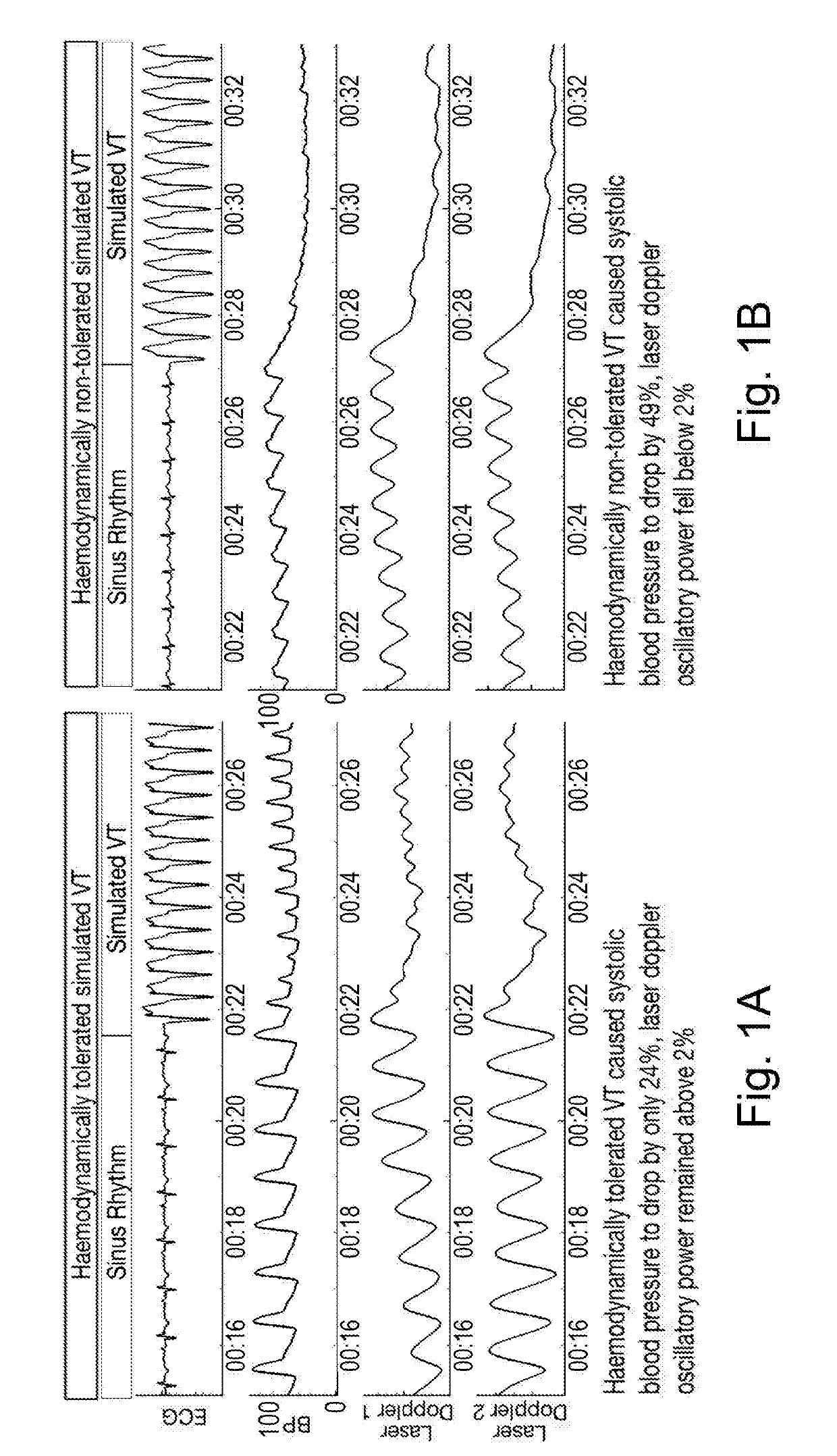
[0064]Many current implantable or externally wearable devices monitor and / or deliver therapies for treating disorders of the cardiac rhythm based on electrical information obtained from the electrical changes that are sensed from the heart. Such electrical activity of the heart represents the heartbeat, but relying solely on this information may not be sufficient to guide therapy reliably as mentioned above.
[0065]Any misguidance of therapy can be particularly harmful where the device is an implantable or externally wearable cardiac defibrillator device which delivers therapy for treating cardiac arrhythmias, because it involves delivering a rapid sequence of electrical stimuli (overdrive pacing) or larger electric shocks (defibrillation) to the heart in an attempt to restore the heart to a normal rhythm. Overdrive pacing can be harmful because sometimes it causes the tachycardia to deteriorate into a faster tachycardia which can worsen the function of the heart. Electric shocks can ...
PUM
 Abstract
Description
Claims
Application Information