Image guided patient setup for radiotherapy

Active Publication Date: 2019-05-23
BRAINLAB
View PDF0 Cites 2 Cited by
  • Summary
  • Abstract
  • Description
  • Claims
  • Application Information

AI Technical Summary

Benefits of technology

The present invention, which is a new type of medical positioning system for radiotherapy treatment, allows medical personnel to position the patient in a more convenient way before treatment. This is done by acquiring images of the patient's outer contour and checking if they are in the correct position. The system can also determine if there are any small positional deviations that may affect the treatment outcome and can make adjustments accordingly. This makes the treatment procedure easier and more efficient, improving the chances of successful treatment.

Problems solved by technology

The problem which arises in this context is that some medical procedures are planned on the basis of images that show certain anatomical structures of interest, but which have been acquired prior to the actual medical procedure.
This problem particularly arises for treatment of the left breast, as the distance from the radiotherapy target to the heart is comparatively short.

Method used


Image

  • Image guided patient setup for radiotherapy
  • Image guided patient setup for radiotherapy

Examples


Embodiment Construction

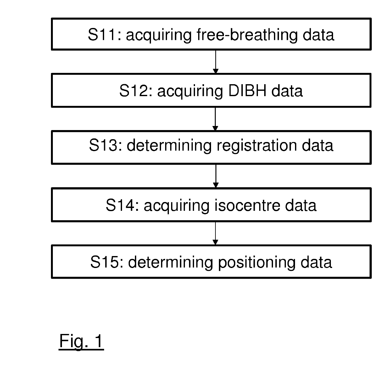
[0061]FIG. 1 illustrates the basic steps of the method according to the first aspect of the present invention. In a first step S11, the spatial position of a reference structure, for example the sternum of a patient is determined for a freely breathing patient. Additionally, the spatial position of that reference structure is determined (step S12), when the patient performs a DIBH, wherein this position can be directly derived from a planning-CT-dataset for which the patient has to perform a DIBH anyhow. The positional data of the reference structure is, if necessary, then registered with the planning-CT-dataset (step S13), which shows for a DIBH-state of the patient not only the pathological target structure (e.g. a breast tumour) to be irradiated, but also the reference structure.

[0062]As soon as the spatial position of the machine isocentre is known (step S14) a spatial position can be calculated (step S15), in which the reference structure of a freely breathing patient has to be...


PUM


Abstract

The present invention relates to a method for setting up a patient for radiotherapy, wherein the patient is scanned prior to therapy in both, a free-breathing state and a DIBH-state for which the patient performs a so-called “deep inspiration breath-hold”. The obtained data is registered to a CT-dataset which forms the basis for a treatment plan. The patient is then set up with respect to a radiation treatment apparatus based on the data obtained for the free-breathing state.

Description

FIELD OF THE INVENTION[0001]The present invention relates to a computer-implemented method for setting up a patient for radiotherapy, a corresponding computer program, a non-transitory program storage medium storing such a program and a computer for executing the program, as well as a medical system comprising an electronic data storage device and the aforementioned computer.TECHNICAL BACKGROUND[0002]For medical procedures performed on a patient, such as image guided surgery, radiotherapy or even image based diagnostics it is desirable to know the spatial position (including the spatial location and / or the spatial orientation), and preferably even the size and the shape of anatomical structures of interest of the patient's body. While some anatomical structures maintain their position over time, some structures do not as they are subject to the patient's respiratory motion. The problem which arises in this context is that some medical procedures are planned on the basis of images th...

Claims


Application Information

Patent Timeline
IPC IPC(8): G06T7/33A61B34/10A61N5/10G16H30/40
CPCG06T7/33A61B34/10A61N5/1049A61N5/1039G16H30/40G06T2207/30096G06T2207/10072A61B2034/107A61B2090/3762G06T2207/10116G06T7/30G06T7/74G06T7/75G06T7/251G06T7/248A61B5/0077A61B5/7292A61N5/1037A61N2005/1059G06T2207/10081G06T2207/20128G06T2207/20221G06T2207/30196G06T7/0014G06T11/003G06T2207/30008G06T2207/30081
Inventor BERLINGER, KAJETANSTEAD, MICHAEL
Owner BRAINLAB
Why Eureka
Try Eureka