
Methods and arrangements for obtaining information and providing analysis for biological tissues

a biological tissue and information technology, applied in the field of biological tissue information and analysis, can solve the problems of not providing biophysical dynamic information about the cornea or lens, ocular analyzer (corneal hysteresis) generally providing only limited information, and no imaging information or data about the lens, etc., to achieve the effect of enhancing brillouin scattering

Pending Publication Date: 2019-11-07
THE GENERAL HOSPITAL CORP
View PDF3 Cites 3 Cited by
  • Summary
  • Abstract
  • Description
  • Claims
  • Application Information

AI Technical Summary

Benefits of technology

This patent describes a technique to characterize tissues in the eye, such as the lens and cornea, using infrared light and elasticity imaging. This method can provide quantitative information about the biomechanical properties of the eye, which can help in diagnosing and screening for various eye diseases. The technique can also combine multiple contrast mechanisms, such as reflectance microscopy, to get a complete characterization of the eye's structure, elasticity, chemical composition, and functional abilities. Additionally, the patent discusses enhancing the Brillouin scattering by using multiple pump beams, auxiliary ultrasound, or contrast-enhancing nanoparticles. Overall, this patent presents a novel and non-invasive technique for in vivo tissue characterization with high resolution.

Problems solved by technology

Other imaging devices, such as the Pentacam (rotating Schiemflug imaging), generally provide only static information about the lens and cornea and likely do not provide biophysical dynamic information about the cornea or lens.
Likewise, the Ocular Analyzer (corneal hysteresis) generally provides only limited information about the cornea regarding its elasticity, and no imaging information or data about the lens.

Method used


Image

  • Methods and arrangements for obtaining information and providing analysis for biological tissues
  • Methods and arrangements for obtaining information and providing analysis for biological tissues
  • Methods and arrangements for obtaining information and providing analysis for biological tissues

Examples


Embodiment Construction

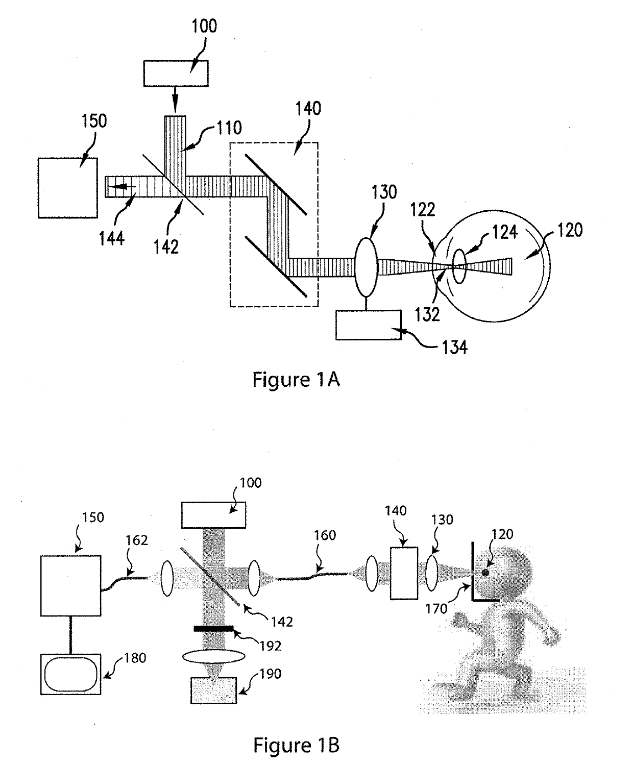
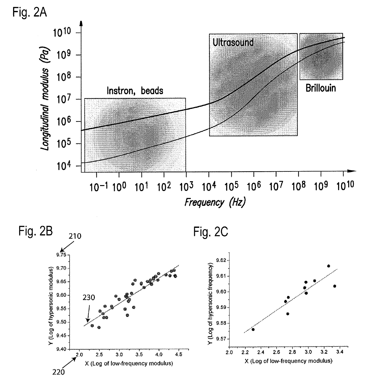
[0070]According to an exemplary embodiment of the system, method and apparatus according to the present disclosure, it is possible to perform a Brillouin microscopy in ocular tissue in vivo, which can be valuable in ocular biomechanical characterization in diagnosing and treating ocular problems, as well as developing novel drugs or treatments.

[0071]There are four anatomical sites in the eye: For example, the cornea is a thin (e.g., less than 1 mm) tissue composed by different layers ofvarying mechanical strength. The aqueous humor is a liquid with similar properties to water that fills the anterior chamber of the eye. The crystalline lens is a double-convex sphere composed by many layers of different index of refraction, density and stiffness. The vitreous humor is the viscous transparent liquid that fills the posterior chamber of the eye.

[0072]Brillouin light scattering in a tissue or any other medium usually arises due to the interaction between an incident light and acoustic wav...


PUM


Abstract

Arrangements and methods can be provided for determining information associated with at least one section of at least one biological tissue. For example, it is possible to provide at least one first electro-magnetic radiation to the section(s) of the biological tissue(s) in vivo so as to interact with at least one acoustic wave in the biological tissue(s). At least one second electro-magnetic radiation can be produced based on the interaction. At least one portion of the at least one second electro-magnetic radiation can be received, and the information associated with the section(s) of the biological tissue(s) can be determined based on the portion of the second electromagnetic radiation(s).

Description

CROSS-REFERENCE TO RELATED APPLICATION(S)[0001]This application is a continuation-in-part of U.S. patent application Ser. No. 13 / 460,595, filed Apr. 30, 2012, which is based upon and claims the benefit of priority from U.S. Patent Application Ser. No. 61 / 480,885, filed Apr. 29, 2011, the entire content of each of which is incorporated herein by reference.FIELD OF THE DISCLOSURE[0002]The present disclosure relates to exemplary methods and apparatus for obtaining information and providing analysis for biological tissues, and more particularly to exemplary methods and apparatus for achieving non-invasive in vivo biomechanical and biophysical characterization, including but not limited to elasticity and viscosity, of various biological tissues, including human and animal eye via, e.g., fast optical spectroscopy and microscopy based on Brillouin light scattering.BACKGROUND INFORMATION[0003]Ectasia is also one of the rare but serious adverse outcomes after LASIK (laser-assisted in situ ke...

Claims


Application Information

Patent Timeline
Patent Type & Authority Applications(United States)
IPC IPC(8): A61B3/107A61B3/10A61B5/00
CPCA61B5/0075A61B3/0008A61F9/008A61B3/102G01J3/4412A61B3/1173A61B5/0066A61B3/107G01J3/0218G01J3/44G01J3/26G01J3/0229G01J3/0208G01J3/10G01J3/2803G01J2003/1213G01J3/0297A61B3/117
Inventor YUN, SEOK-HYUNSCARCELLI, GIULIANO
Owner THE GENERAL HOSPITAL CORP
Learn More