
Treatment and detection of trypanosomes

a trypanosome and tryptosome technology, applied in the field of treatment and detection of trypanosomes, can solve the problems of animal death, significant economic repercussions, and development of effective vaccine strategies, and achieve the effects of effective protection against infection, and effective methods and compositions

Inactive Publication Date: 2019-12-26
UNIV DE BORDEAUX +2
View PDF0 Cites 0 Cited by
  • Summary
  • Abstract
  • Description
  • Claims
  • Application Information

AI Technical Summary

Benefits of technology

This patent is about methods and compositions for treating and diagnosing trypanosome infection. It involves using excreted / secreted antigens (exoantigens) from trypanosomes to identify a protein that can be neutralized or inhibited to protect against infection. This approach can be applied to different strains of trypanosomes, making it an effective way to control infections and treat trypanosomiasis in animals. The invention also allows for the detection and monitoring of trypanosomes and the construction of primers and probes for molecular biology techniques.

Problems solved by technology

In animals, infection causes trypanosomiasis (sometimes called trypanosomosis), which can cause the animal to die.
The subspecies T. evansi is transmitted to cattle, horses and dromedaries, and has significant economic repercussions throughout the cattle-rearing regions.
In infected mammals, the ability of trypanosomes to escape the host's immune defenses by expressing variable antigens on their surface has to date prevented the development of effective vaccine strategies.
Only a few trypanocidal molecules are available, but they cause significant side effects and many resistant parasite strains have appeared.
But there are to date no reliable markers allowing rapid and specific detection of infection, at a reasonable cost.

Method used


Image

  • Treatment and detection of trypanosomes
  • Treatment and detection of trypanosomes
  • Treatment and detection of trypanosomes

Examples


example 1

n of Parasite Growth by a Monoclonal Antibody Against Kinesin

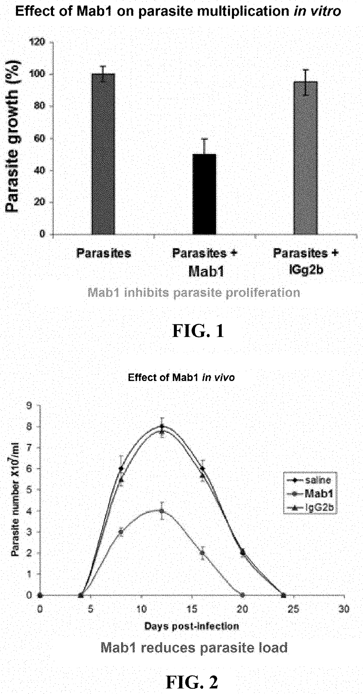
[0133]In vitro, monoclonal antibody Mab1 was added to parasites in co-culture with feeder layers (Mab1 concentration in the culture: 4 μg / mL). Compared with the control, Mab1 inhibits parasite growth whereas the IgG2b isotype control (concentration: 4 μg / mL) has no effect (FIG. 1).

[0134]In vivo, injecting Mab1 (200 μg in 200 μL of PBS intraperitoneally) into mice parasitized for 2 days inhibits the development of parasitemia (expressed in log 10 of parasite number per mL of blood); the IgG2b isotype control (200 μg in 200 μL of PBS intraperitoneally) has no effect (FIG. 2).

example 2

rotection Against Parasites by Vaccination

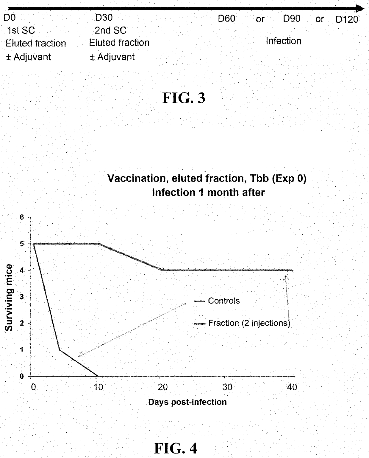
[0135]The fraction enriched in TbKHC1 protein is injected (10-20 μg / mouse) twice, with a 30-day interval (D0 and D30), into the mouse via the subcutaneous route, with or without adjuvant (saponin, 25 μg per mouse). Control mice receive medium alone with or without adjuvant.

[0136]The mice are infected 1, 2 or 3 months after the last injection (see FIG. 3). The parasitemia of the vaccinated mice and of the controls (medium alone±adjuvant) is evaluated daily for 25 days post-infection, then once per week thereafter. The results are presented in FIG. 4.

[0137]Control mice having received the medium with or without adjuvant die around the 7th-8th day post-infection. Remarkably, 22 of 26 mice (84.6%) having received two injections of TbKHC1 protein+adjuvant survive. Adding the adjuvant to TbKHC1 protein increases the survival rate of vaccinated mice and the duration of efficacy of the vaccine.

example 3

on with TbKHC1 Induces Cross-Protection

[0138]Mice are immunized with TbKHC1 protein and then infected 2 months after either with the same trypanosome or with a trypanosome of another species.

Infection 2Number ofmonths afterOrigin ofsubcutaneousthe lastTbKHC1 forinjections +injection +ParasitemiaBatchesimmunizationsaponinadjuvantand survivalBatch 1T. brucei2T. b.No parasite(14 mice)gambiensegambiensedetectedin themice; allsurvive at50 daysBatch 2T. brucei2T. b. bruceiNo parasite(14 mice)gambiensedetectedin themice; allsurvive at50 days

These results show cross-protection, wherein TbKHC1 protein of T. brucei gambiense is capable of inducing protection against infection with T. brucei brucei.


PUM

PropertyMeasurementUnit
molecular weightsaaaaaaaaaa
molecular weightaaaaaaaaaa

Abstract

The present invention relates to methods and compositions for preventing, treating and diagnosing infection by trypanosomes. The invention also relates to the use of excreted / secreted antigens (exoantigens, secretome) and specifically to the identification of a protein excreted / secreted by the trypanosomes, the inhibition of which makes it possible to provide effective protection, mainly by vaccination, against infection by trypanosomes or the development or spread thereof. The invention relates to use of the protein, the derivatives thereof, a nucleotide sequence derived from said protein, or an extract enriched with said protein, and to the use of antibodies directed against said trypanosomes for immunotherapy, diagnosis, and monitoring of infections by trypanosomes.

Description

CROSS-REFERENCE TO RELATED APPLICATIONS[0001]This application is a continuation of U.S. Ser. No. 15 / 575,047, filed Nov. 17, 2017, which is the U.S. national stage application of International Patent Application No. PCT / FR2016 / 051170, filed May 18, 2016.[0002]The Sequence Listing for this application is labeled “Seq-List.txt” which was created on Nov. 15, 2017, and is 52 KB. The entire content of the sequence listing is incorporated herein by reference in its entirety.[0003]The present invention relates to methods and compositions for preventing, treating and diagnosing trypanosome infection. In particular, it relates to the use of excreted / secreted antigens (exoantigens, secretome) and, more particularly, to the identification of a protein excreted / secreted by trypanosomes, the neutralization or inhibition of which makes it possible to confer effective protection against infection with trypanosomes or the development or spread thereof, mainly by vaccination. The invention enables a ...

Claims


Application Information

Patent Timeline
Patent Type & Authority Applications(United States)
IPC IPC(8): A61K39/005A61P33/02C07K16/20G01N33/569
CPCG01N2800/26G01N33/56905G01N2800/52A61K39/005A61P33/02C07K16/20C07K2317/76A61K38/16A61K31/7105C07K14/44A61K2039/55577A61K2039/58A61K2039/505
Inventor VINCENDEAU, PHILIPPELEMESRE, JEAN-LOUPPAYS, ETIENNE
Owner UNIV DE BORDEAUX
Learn More