Renovascular treatment device, system, and method for radiosurgically alleviating hypertension

a treatment device and a technology for hypertension, applied in the field of improved devices, systems and methods for treating patients, can solve the problems of affecting the treatment effect, affecting the patient's comfort, so as to avoid potential implant-induced occlusive response, improve patient comfort, and avoid the effect of reducing the risk of complications

Active Publication Date: 2020-01-16
CYBERHEART
View PDF0 Cites 0 Cited by
  • Summary
  • Abstract
  • Description
  • Claims
  • Application Information

AI Technical Summary

Benefits of technology

[0010]The renal nerves, owing to their position vis-à-vis the renal arteries, may correspond to moving tissue of a patient, both as a result of respiration of the patient, the pulsation of the aorta and renal artery, and the heartbeat of the patient. With respect to the latter, the temporal rise and fall of blood pressure of the heartbeat cycle may cause the outer diameter of the renal arteries to expand and contract by an amount that is sufficient to move the renal nerves by an amount which may affect the treatment. Embodiments of the invention allow improved radiosurgical treatments of tissues of the renovascular system, often enhancing the capabilities of existing radiosurgical systems for targeting what in at least some instances may be relatively rapidly moving tissues (renal nerves) so as to mitigate hypertension. Treatment of renal nerves may benefit from an implanted position surrogate for identification of the location of the target tissue, with the surrogate optionally comprising a fiducial marker positioned in or near the renal arteries and / or the kidneys using catheterization techniques or direct transcutaneous routes. Novel catheters and / or delivery structures having active fiducials may limit the need for X-rays (and thereby minimize collateral imaging radiation exposure). Enhanced planning and tracking techniques may also be employed, with the radiosurgical renovascular system treatments described herein generally being compatible with many components of existing radiosurgical treatment systems.
[0014]In many embodiments, implanting of the surrogates will comprise advancing at least one elongate flexible body through a blood vessel. The surrogate may be coupled to tissue so that the surrogate exhibits heartbeat- and / or respiratory-induced movement. The implanted surrogate may comprise a non-colinear set of discrete fiducial markers so that a three-dimensional offset orientation between the surrogate and the target area can be determined from an image of the fiducial markers. In some embodiments, implanting of the surrogate may comprise screwing a helical structure of the elongate body into a soft, contractile tissue of the heart. Implanting of the surrogate may also include expanding an expandable body with a lumen or cavity bordered by the tissue, with the expandable body optionally comprising an inflatable balloon, a temporary stent-like structure, or the like, which can be safely and reversibly expanded within a vessel so as to engage the surrounding tissue. In exemplary embodiments, implanting the surrogate may comprise fixing an active three-dimensional position indicator to the tissue, with the position indicator transmitting a position indicating signal that can be used to register a location of the implanted surrogate with the planning image data. In many embodiments, the fiducial(s) will be implanted prior to acquiring planning image data. In other embodiments, an image taken after implanting the surrogate may facilitate registration. For example, when the position indicating signal indicates an offset between the surrogate and a position sensor (or transmitter) disposed outside the body, the position indicating signal can be calibrated using post-implant image data that encompasses the position sensor. In one exemplary embodiment, the image data used for calibrating the position indicating signal comprises post-planning calibration image data, and a calibration position sensing signal is generated while a catheter tip engages a heart tissue. A positional relationship between the sensor and the body is maintained during acquisition of the calibration image data and the generation of the position sensing signal. More generally, the position indicator typically comprises a sensor or signal generator used within ultrasound or electromagnetic position indicating systems. The target region can be treated by directing the planned radiation using a position indicating signal from the position indicator between intermittent tracking verification images. Hence, position surrogates employing such active fiducial systems may limit the need for imaging X-rays (and thereby minimize collateral imaging radiation exposure).
[0026]According to another aspect of the present invention, there is a radiosurgical system for treating a patient body with a renal artery and hypertension. The system comprises an image capture device for acquiring three dimensional planning image data from the renal artery and / or a location proximate the renal artery, a radiation source for transmitting a plurality of beams of ionizing radiation from outside the body, and a processor system configured to direct the ionizing radiation beams toward a target region of the renal artery and / or a target region at the location proximate the renal artery such that the radiation beams remodel the target region and the hypertension is mitigated.
[0027]In some embodiments, a position surrogate may be positioned and / or implanted from within an inferior vena cava (IVC) of the body prior to acquiring of the planning image data. The IVC position surrogate may be temporary, but will often remain implanted at least throughout planning and radiation delivery (optionally being permanent). Advantageously, an IVC surrogate may comprise a bioresorbable or biodegradable structure. Any inadvertent emboli associated with an IVC position surrogate may be relatively safely directed to the pulmonary vasculature by the bloodflow, and access to the IVC for implantation of prosthetic structures is well established with good patient safety. Surprisingly, in many of the embodiments described herein (including those employing an IVC position surrogate), no position surrogate may be implanted within at least the first renal artery, often within either of the renal arteries. This lack of a surrogate within the renal artery may help avoid challenges of accessing the renal arteries, and perhaps more importantly, may completely avoid potential implant induced-occlusive response of the tissues of the renal arteries (which could otherwise induce associated increase in blood pressure and thereby potential decrease or even overwhelm the hypertension alleviation available via denervation).

Problems solved by technology

Ablating the origin of the renal nerves in the sympathetic ganglia has historically been considered very difficult.
Pharmacologic assault on nerve functions is associated with systemic complications.
The sympathetic renal nerves arborize throughout the walls of the renal arteries, and frustrate access thereto.
Unfortunately, some radiosurgical therapies, and particularly those which seek to target and track moving tissues using x-ray imaging, fluoroscopy, or other remote imaging modalities, may subject collateral tissues to significant imaging-related radiation and associated injury.

Method used


Image

  • Renovascular treatment device, system, and method for radiosurgically alleviating hypertension
  • Renovascular treatment device, system, and method for radiosurgically alleviating hypertension
  • Renovascular treatment device, system, and method for radiosurgically alleviating hypertension

Examples


Embodiment Construction

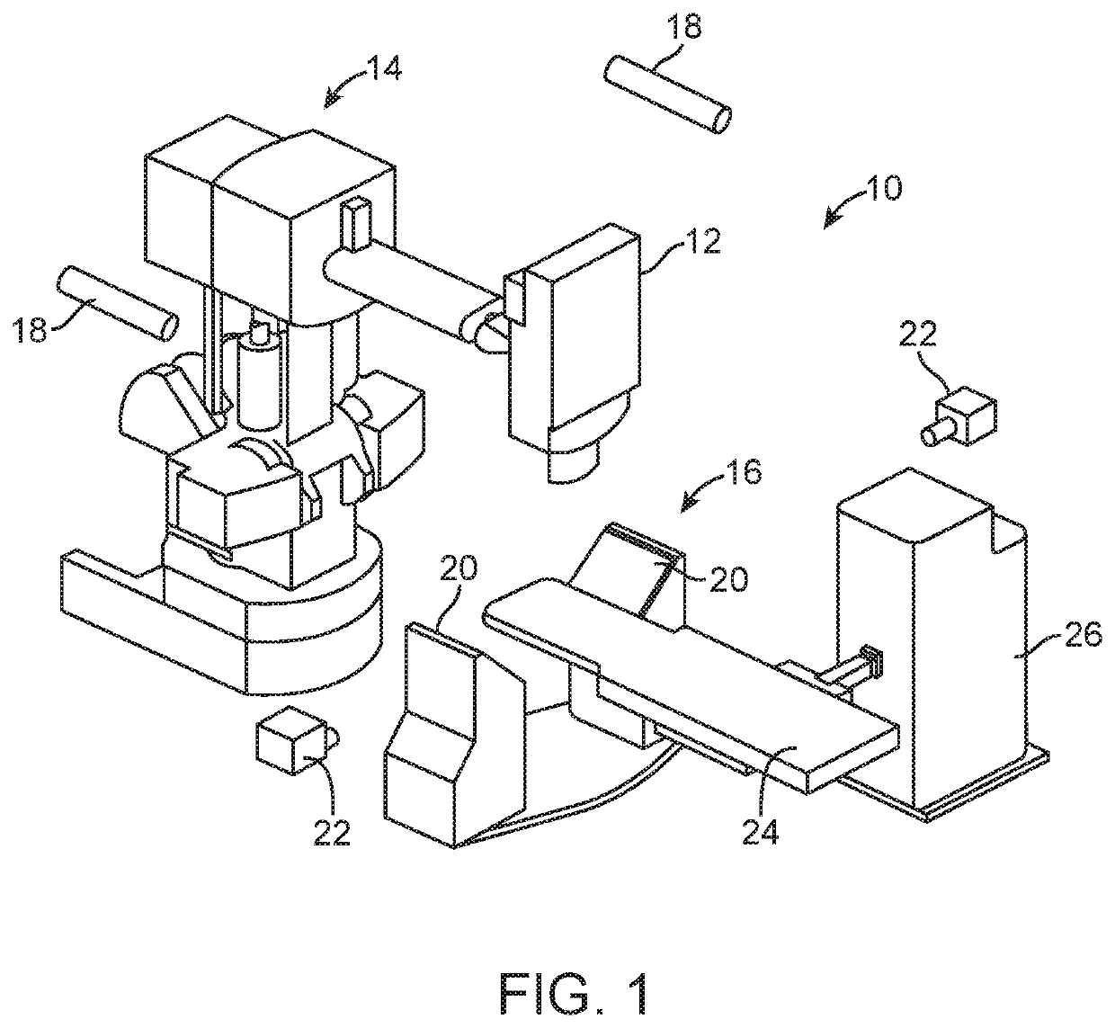
[0062]The present invention generally provides improved systems, devices, and methods for treatment of tissue to alleviate cardiorenal disease, including hypertension, using radiosurgical systems. The embodiments of the present invention detailed herein and variations thereof may have the therapeutic effect of slowing, including halting, the progression of congestive heart failure via a non-invasive or minimally invasive (in embodiments utilizing catheters or percutaneous needles to implant surrogates, as detailed below) surgical procedure. Some or all embodiments detailed herein and variations thereof may be practiced to mitigate the conjunctive heart failure that sometimes develops following a myocardial infarction.

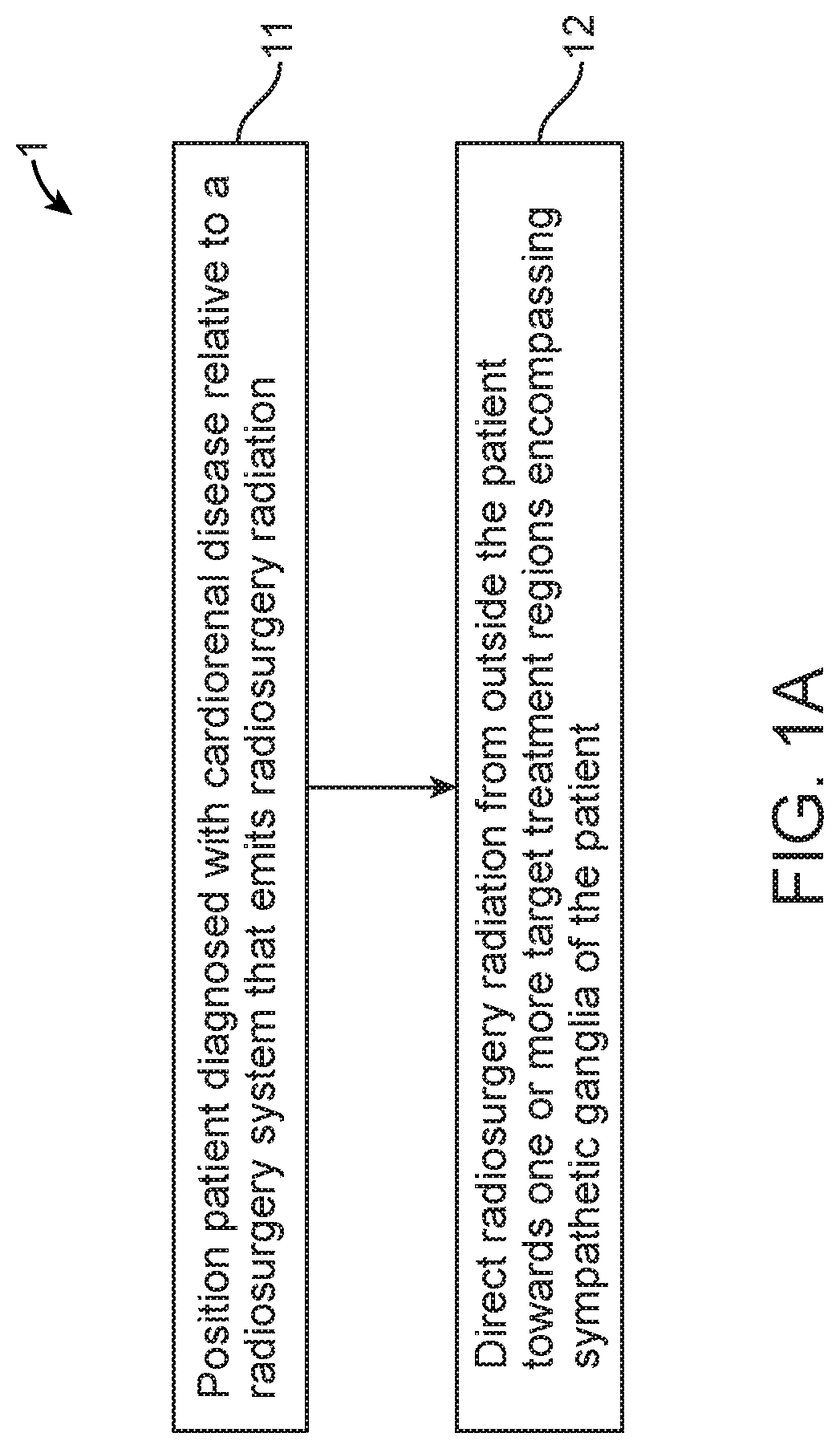
[0063]FIG. 1A provides an exemplary flowchart 1 of a method for treating cardiorenal disease of a patient, such as hypertension. At step 11, a patient diagnosed with cardiorenal disease, such as hypertension, is positioned relative to a radiosurgery system such as that ...


PUM


Abstract

A radiosurgical method for treating cardiorenal disease of a patient, the method including directing radiosurgery radiation from outside the patient towards one or more target treatment regions encompassing sympathetic ganglia of the patient so as to inhibit the cardiorenal disease. In an exemplary embodiment, the method further includes acquiring three dimensional planning image data encompassing the first and second renal arteries, planning an ionizing radiation treatment of first and second target regions using the three dimensional planning image data so as to mitigate the hypertension, the first and second target regions encompassing neural tissue of or proximate to the first and second renal arteries, respectively, and remodeling the target regions by directing the planned radiation from outside the body toward the target regions.

Description

CROSS REFERENCE TO RELATED APPLICATION DATA[0001]The present application claims the benefit under 35 USC 119(e) of U.S. Provisional Application No. 61 / 483,962 filed May 9, 2011. The full disclosure of which is incorporated herein by reference in its entirety for all purposes.BACKGROUND OF THE INVENTION[0002]The present invention generally provides improved devices, systems, and methods for treatment of a patient. Exemplary embodiments provide radiosurgical treatment of tissues, including nerves, in a patient body, often to treat a renovascular disease and / or to treat heart failure. Exemplary embodiments may deposit a sufficient ionizing radiation dose at a target region of the renovascular system in general, and renal nerves located proximate to the renal arteries in particular, so as to treat hypertension of the patient body. Along with allowing treatment of tissues which may move at a relatively rapid pace, embodiments of the invention may accommodate significant deformation or re...

Claims


Application Information

Patent Timeline
Patent Type & Authority Applications(United States)
IPC IPC(8): A61N5/10
CPCA61N5/1039A61B2018/00404A61B2018/00434A61B2018/00511A61N5/1042A61N5/1065A61N5/1083A61N5/1084A61N2005/1061
Inventor MAGUIRE, PATRICKGARDNER, EDWARD
Owner CYBERHEART
Why Eureka
Try Eureka