Patient specific 3-d interactive total joint model and surgical planning system

a total joint model and patient technology, applied in the direction of shoulder joints, prostheses, manufacturing tools, etc., can solve the problems of inadequate bone affixing, inability to satisfactorily determine the orientation of a joint replacement, and wear of bones, so as to reduce the penetration of bone, confirm the configuration of bone fixation, and increase the tuberosity

Active Publication Date: 2020-01-30
NEW YORK SOC FOR THE RUPTURED & CRIPPLED MAINTAINING THE HOSPITAL FOR SPECIAL SURGERY
View PDF0 Cites 7 Cited by
  • Summary
  • Abstract
  • Description
  • Claims
  • Application Information

AI Technical Summary

Benefits of technology

[0010]This and other embodiments can include one or more of the following features. The method of conducting pre-operative planning can include one or more of analyzing a joint line, including comparing an original joint line and a new joint line, wherein the new joint line is substantially similar to the original joint line. The method of the step of conducting pre-operative planning can include comparing vectors in three dimensions which represent the distance and direction between tendon and muscle insertions on the scapula and the humerus for measuring the distance of relocation of humeral tuberosity compared to the scapula; determining the diameter of the humeral head; determining the height of humeral head; determining the size of humeral bone implant from digital images. The method of the step of determining a best fit size of a joint implant can include selecting a humeral implant from a range of sizes, wherein the range of sizes is selected from the group consisting of length of stem, size of humeral stem, diameter of stem, size diameter of head, height of head, and offset of the center spherical head compared to the center of the face of the humeral stem. The method of the step of conducting range of motion analysis can include simulating motion of the virtually implanted joint through extreme ranges of motion to measure impact locations and compensate for necessary functional range of motion. The method of the step of conducting soft tissue analysis can further include: determining key soft tissue insertion points, measuring distances in three dimensions for comparison to pre-operative conditions, and assessing lengths at extreme ranges of motion, such that total soft tissue length change or contraction is substantially maintained within anatomical ranges in order to substantially achieve most common activities of daily living. The method of the step of assessing and adjusting the characteristics of the joint implant can include: assessing and adjusting the thickness/height of the glenoid implant; assessing and adjusting the depth of the glenoid fossa; and assessing and adjusting the thickness of a graft. The method of the step of selecting patient specific instruments can include selecting a humeral implant and a glenoid implant based on the pre-operative analysis, or assessing and adjusting a humeral head, a glenoid thickness, a glenoid fossa depth, and a graft thickness based on the pre-operative analysis. The method can further include instructions for a comprehensive pre-operative total joint planning method for comparison of anatomic or reverse shoulder surgery implants or guides. The method can further include conducting range of motion analysis, including virtually positioning implants through extreme ranges of motion to measure impact locations and compensate for necessary functional range of motion. The method can further include conducting soft tissue analysis, including determining key soft tissue insertion points, measuring distances in three dimensions for comparison to pre-operative conditions, and assessing lengths at extreme ranges of motion, such that total soft tissue length change or contraction is substantially maintained within anatomical ranges in order to substantially achieve most common activities of daily living. The method can further include assessing and adjusting as needed the thickness/height of the glenoid implant. The method can further include assessing and adjusting as needed the depth of the glenoid fossa. The method can further include assessing and adjusting the thickness of a graft. The pre-operative planning can be done virtually based on images taken from a subject prior to surgery. The method can further include optimizing the dimensions of fixation elements of the glenoid implant using correspondence matrix between a three dimensional (3D) bony structure of the patient and a statistical shape based atlas according to the following steps: (1) developing a registration between patient bone and statistical shape model of the bone of interest; (2) extracting the principle modes representing the patient bone; (3) defining the fixation configuration, position or dimensions according to the corresponding modes; and (4) applying collision detection to confirm the configuration of the bone fixation. The method can further include identifying and comparing procedural risks between selected reverse or anatomic shoulder procedures by determining: whether a glenoid face coverage is maximized; whether an overhang of the glenoid face is minimized; whether bone removal on the glenoid face is minimized; whether the glenoid retroversion is less than about 5 to about 10 degrees; whether seating of the glenoid implant is greater than about 80% of the implant coverage area; whether there is minimized penetration of the glenoid cortical wall anteriorily; whether there is greater than about 3 mm bone thickness behind glenoid; whether the orientation offset between the native glenoid and implant superior/inferior axis is less than about 5 degrees; whether the superior or inferior tilt versus native glenoid is less than 5 degrees; whether there is an absence of a humeral head overhang compared to the cut, or prepared surface of the humeral bone; whether there is less than about 3 mm difference in humeral head diameter between anatomic and implant; whether there is less than about 1 mm difference in humeral head height between anatomic and implant; and whether there is less than about 2 mm greater tuberosity to medial head edge in comparison to anatomic; whereby procedural risks are identified; and the selection of prosthetic implants for the selected shoulder surgery are based at least in part on the identified procedural risks. The glenoid implant can be augmented to fit a patient for which pre-operative planning of an anatomic or reverse shoulder procedure was performed. The depth of augmentation, the size of augmentation, and/or the radial position of augmentation can vary depending on the pre-operative planning of a selected reverse or anatomic shoulder procedure. The augmentation can include a depth ranging from about 2 mm to about 4 mm. The augmentation can cover about 5%, 10%, 15%, 20%, 30%, 40% or 50% of the back side of the glenoid implant. The augmentation can covers about 50%, 60%, 70%, 80%, 90%, 95% or greater of the back side of the glenoid implant. The radial location of the augmentation on the backside of the glenoid implant can be selected from the group consisting of a posterior location, an anterior location, a superior location, an inferior location, and combinations thereof. The method can further include obtaining a patient specific shoulder surgery guide based upon the selected anatomic or reverse surgical method steps. The method can further include producing a shoulder surgery guide or a selected reverse or anatomic shoulder procedure, wherein producing the shoulder surgery guide comprises using a 3D printing device. The method can further include a selected anatomic or reverse shoulder procedure recommending prosthetic shoulder implants and placement positions, selected from the group consisting of adjustments in glenoid implant size, augmentation depth, augment position, positioning in six degrees of freedom, fixation type, fixation size, reaming depth, reaming diameter, reaming angle(s) and/or a combination thereof. The method can further include a computer readable medium having stored thereon executable instructions that when executed by the processor of a computer control the

Problems solved by technology

In some patients, however, one or more of the bones of the shoulder are not only arthritic, but have also had previous conditions that have caused bone to wear away.
In such cases, there may not be sufficient bone to adequately affix a prosthetic implant to the bone, or the bones may have been worn such that the orientation of a joint replacement cannot be satisfactorily determi

Method used


Image

  • Patient specific 3-d interactive total joint model and surgical planning system
  • Patient specific 3-d interactive total joint model and surgical planning system
  • Patient specific 3-d interactive total joint model and surgical planning system

Examples


Embodiment Construction

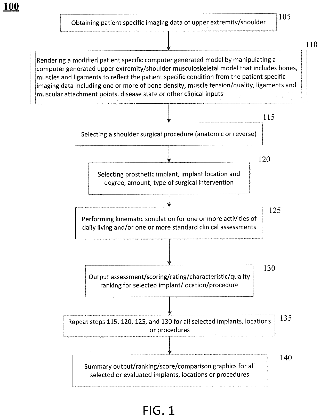
[0029]Patients requiring shoulder surgery may have one or more of the bones of the shoulder that are not only arthritic, but may also have had previous conditions that have caused bone to wear away. In such cases, there may not be sufficient bone to adequately affix a prosthetic implant to the bone during a routine shoulder surgery. Indeed, the bones may have been worn such that the orientation of a joint replacement cannot be satisfactorily determined to ensure a positive patient outcome.

[0030]The glenoid bone can be subject to increased wear due to bone arthritic conditions of the joint, and due to alterations of a normal soft tissue envelope surrounding the joint. In such cases, the orientation of the face of the glenoid portion of the scapula bone may be altered so that the humeral bone is no longer appropriately opposed to the glenoid surface. In the case where the glenoid is severely worn, there can be two or more risks a surgeon must balance in an attempt to improve shoulder ...


PUM

PropertyMeasurementUnit
Fractionaaaaaaaaaa
Fractionaaaaaaaaaa
Fractionaaaaaaaaaa

Abstract

Methods, systems and devices for pre-operatively planned total or partial joint surgery including, for example, anatomic and reverse shoulder surgery guides and implants. There are also methods for pre-operative planning methods for designing glenoid implants and prostheses, particularly with patient-specific augmentation, based on considerations of multiple factors affecting the outcome of a selected reverse or anatomic shoulder surgery. There are also described methods of performing total or partial joint surgery, including anatomic or reverse shoulder surgery, using surgery guides and implants in patients undergoing joint surgery.

Description

CROSS REFERENCE TO RELATED APPLICATIONS[0001]This application claims priority to U.S. Provisional Patent Application No. 62 / 405,814, filed Oct. 7, 2016, titled “PATIENT SPECIFIC 3-D INTERACTIVE SHOULDER MODEL AND SURGICAL PLANNING SYSTEM,” U.S. Provisional Patent Application No. 62 / 426,081, filed Nov. 23, 2016, titled “PATIENT SPECIFIC 3-D INTERACTIVE SHOULDER MODEL AND SURGICAL PLANNING SYSTEM” each of which is incorporated herein by reference in its entirety.FIELD[0002]The presently disclosed subject matter relates to methods, systems and devices for virtual pre-operatively planned implants and prostheses for use in comparison and planning of total joint arthroplasty. More particularly, shoulder implants and prostheses for use in comparison and planning of anatomic and reverse shoulder procedures are described. In addition, the presently disclosed subject matter also relates to the use of such implants and prostheses in patients undergoing a selected and planned partial or total j...

Claims


Application Information

Patent Timeline
IPC IPC(8): A61B34/10A61F2/46A61F2/30A61F2/40
CPCA61F2/3094A61F2002/30952A61B2034/108A61F2/4081A61B34/10A61F2/46A61F2002/30985A61B2034/102A61F2/40
Inventor KONTAXIS, ANDREASGULOTTA, LAWRENCE
Owner NEW YORK SOC FOR THE RUPTURED & CRIPPLED MAINTAINING THE HOSPITAL FOR SPECIAL SURGERY
Why Eureka
Try Eureka