Modulated ultra-sound compatible artificial cranial prosthesis

a cranial prosthesis and ultra-sound technology, applied in the field of ultra-sound compatible artificial cranial prosthesis, can solve the problems of difficult to determine the efficacy of current imaging techniques (e.g. computed tomography or magnetic resonance imaging) in real time, patients may not respond to certain procedures and/or adjuvant therapies, and achieve the effect of facilitating therapeutic ultrasound application, high intensity focused ultrasound treatment, and effective use of ultrasound

Active Publication Date: 2020-01-30
SAMPATH PRAKASH +1
View PDF0 Cites 6 Cited by
  • Summary
  • Abstract
  • Description
  • Claims
  • Application Information

AI Technical Summary

Benefits of technology

[0008]The aim of the present invention is to enable an effective use of ultrasound in cerebral diagnostics and to further use the claimed device as a platform onto which other diagnostic, delivery, and / or therapeutic devices may be launched, so as to make it possible to perform complete, post-operative imaging of the intracranial contents in real time. In addition, the device will have modulations that can monitor the progress of an intracranial pathology, as well as utilizing the device to administer and deliver therapies when necessary. Finally the device would facilitate therapeutic ultrasound application including blood brain barrier disruption, blood clot liquefication, and high intensity focused ultrasound treatment for brain lesioning.

Problems solved by technology

Such therapies, however, can have marked side effects and it is often difficult to determine their efficacy with current imaging techniques (e.g. Computed Tomography or Magnetic Resonance Imaging) in real time.
Furthermore, some patients may not respond to a certain procedures and / or adjuvant therapies in a timely manner necessitating the need to continually monitor and identify such cases and apply different treatments.
Although ultrasound is a widely-used tool in the field of general diagnostic radiology, it is limited to very few areas in cerebral diagnostics.
Repositioning or replacing the bone flap, removed following the neurosurgical procedure, in fact constitutes a barrier to ultrasound penetration and does not allow follow-up imaging of the patient using ultrasound.
Although occasionally available, the ultrasound methods used to get past the calvarium / skull and / or prosthesis still do not enable an accurate and definite evaluation of the intracranial contents including the brain parenchyma and ventricles.

Method used


Image

  • Modulated ultra-sound compatible artificial cranial prosthesis
  • Modulated ultra-sound compatible artificial cranial prosthesis
  • Modulated ultra-sound compatible artificial cranial prosthesis

Examples


second embodiment

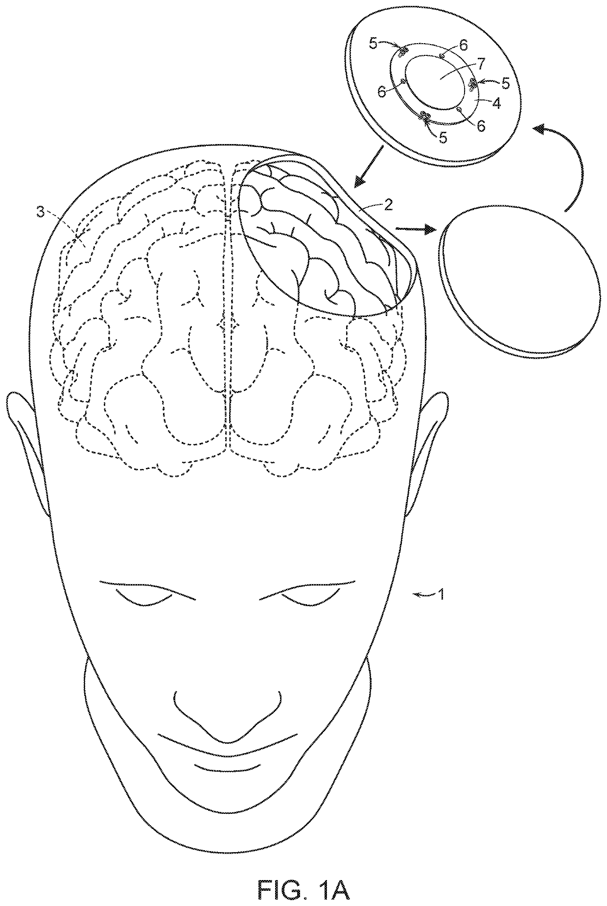
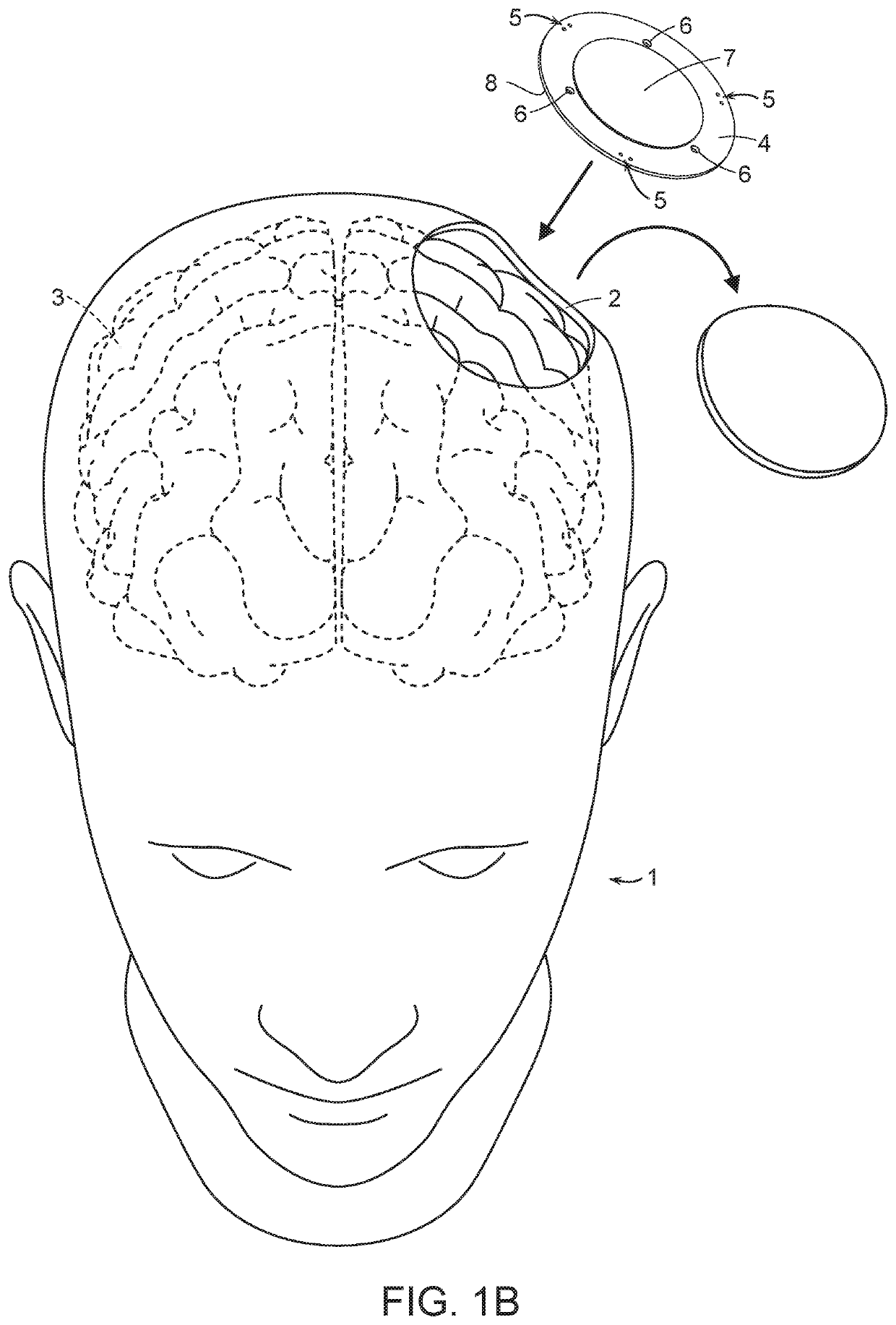
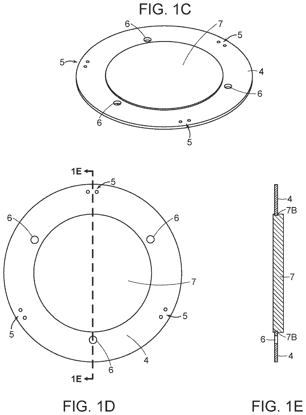
[0034]The cranial prosthesis FIGS. 1C-1E according to the present invention is comprised of an inner, ultra-sound compatible disc 7 surrounded by an outer, rigid ring. The outer rigid ring 4 is configured to accept, i.e. “modulate”, a variety of diagnostic tools 10-12 and devices to monitor various conditions of the brain whilst the treating surgeon uses ultrasound technology to image the brain from the inner disc 7. In a second embodiment FIGS. 2A-2C and FIG. 3, the inner disc 7 is formed with a circumferential groove 7A to accept a circular flange 4A of the inner circumference of the outer ring 4 securing the inner disc 7 to the outer ring 4 by a “press fit.” In yet another embodiment FIGS. 4A-4D and FIGS. 5A-5D, the device further comprises an outer casing 8 which allows for the rotation of the outer ring 4. The rotation of the outer ring 4 allows for the positioning or re-positioning (in the event of re-operation) of the access ports 6 found in said ring 4 at the desired site in...

first embodiment

[0039]In the instant invention FIGS. 1C-1E, the cranial prosthesis is comprised of an inner radio-lucent disc 7 having a planar or curved body unitarily formed from a single piece of material that can allow the use of ultra-sound diagnostic instruments. The radio-lucent section 7 should be constructed so as to prevent the creation of artifacts and / or causing visual impairment. Surrounding the radio-lucent section is an outer modulation ring 4 having a plurality of access ports 6 capable of introducing one or more diagnostic instruments or delivery vehicles 10-12, i.e. “a module”, into the brain of the patient. Said diagnostic instruments 10-12 may be integrated into said outer modulation ring 4 of the cranial prosthesis, either permanently or on a need basis, so that said module is operative while engaged with the cranial prosthesis. The instant device is designed in such a way as to allow for the ultrasound imaging of the brain of the patient while the modulated diagnostic instrume...

third embodiment

[0041]In the claimed invention FIGS. 4A-4D and FIGS. 5A-5D, the inner, ultrasound compatible disc 7 is permanently secured to the outer, modulation ring 4. The outer, modulation ring 4 with the inner ultrasound compatible disc 7, fit within an outer casing 8 having an inner space 8A which receives the outer modulation ring 4 / inner disc 7. The outer casing 8 includes means 9 in which to secure the device to the remaining cranium 2 of the patient 1. Once implanted onto the brain 3 of the patient 1, the outer casing 8 secures the modulation ring 4 / inner disc 7 to the patient 1. The outer modulation ring 7 is able to freely rotate within the space 8A of the outer casing 8 providing the practitioner with the ability to position an access port (not shown) over the desired location aided by the ultrasound imaging. The outer casing 8 has means of securing 9 the device to the cranium 2 of the patient 1 using suture thread, braces, screws, plates 9, bone anchors, sutures, wires or other FDA-a...


PUM


Abstract

An intracranial prosthesis comprised of a flat body having an interior ultrasound-compatible window and means about the outer portion capable of engaging a plurality of diagnostic instruments and/or intracranial delivery systems so that a practicing medical professional can monitor certain parameters of a patient or deliver therapeutic agents to the patient while using an ultrasound-monitoring device to image the patient's brain. The prosthesis is designed to allow for the continuous, uninterrupted, simultaneous monitoring of a number of parameters of a patient's brain at the patient's bedside.

Description

CROSS-REFERENCE TO RELATED APPLICATIONS[0001]This application is a continuation under 35 U.S.C. § 371 of International Application No. PCT / US2017 / 053847 filed on Sep. 27, 2017, which claims priority to U.S. Provisional Patent Application Ser. No. 62 / 400,607 filed Sep. 27, 2016, which is hereby incorporated by reference herein in its entirety.BACKGROUND OF THE INVENTION[0002]The present invention relates to an ultrasound-compatible cranial prosthesis, with modular capabilities, used as a “window” into the cranial vault.[0003]Surgical efforts to repair cranial defects commonly occur. For example, various types of surgical procedures on the human brain require removal of a portion of the skull. By way of example only, those include surgeries that remove brain tumors, reduce brain swelling, repair cerebral aneurysms, evacuate hematomas, remove shrapnel and bullets secondary to trauma, drain abscesses and other intracranial infections, address congenital defects of the brain, as well as ...

Claims


Application Information

Patent Timeline
Patent Type & Authority Applications(United States)
IPC IPC(8): A61F2/28A61B8/08A61B5/00A61B8/00
CPCA61B5/026A61F2250/0093A61B10/02A61B5/031A61F2220/0008A61B5/055A61B8/0808A61F2/2875A61B8/4272A61B5/4839A61B5/14553A61B8/4416A61B2560/0443A61B5/686A61B5/01A61B5/6864A61D7/00A61M2039/025A61B50/20A61B50/24A61B17/688A61B5/4064A61N7/02A61B2018/00577A61B2090/103A61N1/0539
Inventor SAMPATH, PRAKASHDIMECO, FRANCESCO
Owner SAMPATH PRAKASH
Why Eureka
Try Eureka