Scattered x-ray detection to detect misuse and prevent harm

a technology of x-ray detection and x-ray imaging, applied in the direction of radiation diagnostic diaphragms, applications, and diaphragms used in the use of diaphragms/collimeters, etc., can solve the problems of user ignorance, x-ray radiation exposure, and incorrect usage of x-ray imaging systems, so as to improve the safety of x-ray imaging system usag

Active Publication Date: 2020-06-18
KONINKLJIJKE PHILIPS NV
View PDF0 Cites 2 Cited by
  • Summary
  • Abstract
  • Description
  • Claims
  • Application Information

AI Technical Summary

Benefits of technology

[0003]There may therefore be a need for solutions that improve usage safety of X-ray imaging systems.
[0011]The proposed apparatus uses the information in the scatter radiation to increase operational safety of X-ray imaging. Scattered X-radiation, usually considered a nuisance, is put to good use with the proposed system to enhance safety. A risk for misuse is reduced and even less qualified personnel may be allowed to perform X-ray imaging. But even experienced staff more often than not required to working long hours in ever busy medical facilities may benefit from the proposed safety features. In embodiments of feature i), the scatter measurements are processed by the predictor component to derive the predictor signal as the second object is approaching the one or more of the sensors.
[0012]According to one embodiment, the apparatus further comprising a transducer configured to issue an alert signal in response to the predictor signal, said transducer being any one or a combination of i) auditory or ii) optical. This allows quickly alerting user and others that the X-ray imager is incorrectly used.
[0017]According to one embodiment, the apparatus the X-ray imaging apparatus comprises an X-ray detector for imaging the first object, and where the at least one sensor is different from said X-ray detector. This allows arranging the sensors (each having preferably a smaller field of view than the detector) in numerous spatial layouts to reliably capture intensity changes of the scattered radiation.
[0018]Alternatively, and according to one embodiment, the at least one sensor is part of the X-ray detector. More particularly, the one or more sensors comprise one or more pixels of the X-ray detector. This embodiment may be realized by a grating structure as will be explained in more detail below. This embodiment allows reducing parts count and easier deployment as the mounting of the sensor can be dispensed with.
[0019]According to one embodiment, operation of the predictor component is based on a machine learning algorithm trained by one or more samples of X-radiation scatter measurements. Neural networks or other machine learning techniques can be used to enhance reliability and robustness of the predictions.

Problems solved by technology

With wide deployment of DXR (diagnostic X-ray) systems, especially in high throughput centers as well as in centers managed by less qualified personnel, hazardous situations may emerge where people other than the patient may be exposed to X-ray radiation if the system is operated in the presence of unwarranted people in unsafe proximity.
This situation may result from a lack of awareness of the user, or from stressful situations in a crowded hospital or through unauthorized / unscrupulous usage.
Other incorrect usages of X-ray system may also be observed in these settings.

Method used


Image

  • Scattered x-ray detection to detect misuse and prevent harm
  • Scattered x-ray detection to detect misuse and prevent harm
  • Scattered x-ray detection to detect misuse and prevent harm

Examples


Embodiment Construction

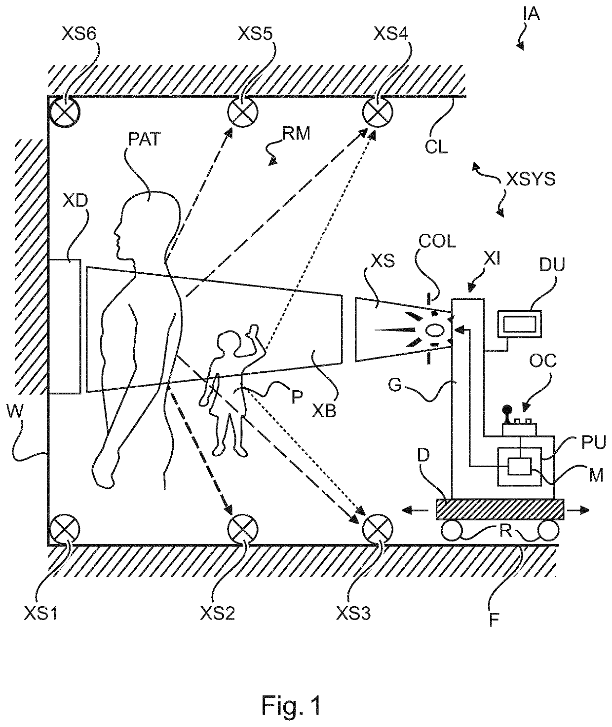
[0052]With reference to FIG. 1, there is shown an imaging arrangement IA. The arrangement includes an X-ray imaging apparatus XI (also referred to herein as the “(X-ray) imager”) and a system XSYS comprising safety module M (referred to herein as “the module”) that supports safe operation of the X-ray imager in a manner to be described in more detail below.

[0053]The X-ray imager XI as mainly envisaged herein is of the mobile or portable type. One such embodiment of a mobile X-ray imager XI is shown schematically on the bottom right of FIG. 1.

[0054]The X-ray imager XI is mobile or portable in the sense that it can be moved from one location, such as from an examination room RM, to another location, in particular to another examination room. This is particularly useful in settings where it is impractical to have a dedicated examination room. The imaging can advantageously be performed in any room that happens to be vacant at a given time. Preferably, there is no disassembling required...


PUM


Abstract

An apparatus and related method for supporting X-ray imaging. The apparatus comprises an input interface (IN) for receiving an X-radiation scatter measurement obtained by an X-ray sensor (SXi) during operation of an X-ray imager (XI) for imaging a first object (PAT). A predictor component (PC) is configured to predict, based on said measurement, whether or not: i) a second object (P) is present, or ii) there is sufficient X-ray exposure of said first object (PAT). The apparatus comprises an output interface (OUT) for outputting a predictor signal indicative of an outcome of said prediction.

Description

FIELD OF THE INVENTION[0001]The invention relates to an apparatus for supporting X-ray imaging, to an imaging system, to a grid structure, to a method of supporting X-ray imaging, to a computer program element, and to a computer readable medium.BACKGROUND OF THE INVENTION[0002]With wide deployment of DXR (diagnostic X-ray) systems, especially in high throughput centers as well as in centers managed by less qualified personnel, hazardous situations may emerge where people other than the patient may be exposed to X-ray radiation if the system is operated in the presence of unwarranted people in unsafe proximity. This situation may result from a lack of awareness of the user, or from stressful situations in a crowded hospital or through unauthorized / unscrupulous usage. Other incorrect usages of X-ray system may also be observed in these settings.SUMMARY OF THE INVENTION[0003]There may therefore be a need for solutions that improve usage safety of X-ray imaging systems.[0004]The object ...

Claims


Application Information

Patent Timeline
Patent Type & Authority Applications(United States)
IPC IPC(8): A61B6/10A61B6/00G21K1/02
CPCA61B6/483A61B6/548G21K1/025A61B6/563A61B6/583A61B6/107A61B6/542A61B6/545A61B6/4291A61B6/4405A61B6/582A61B6/06A61B6/586A61B6/587
Inventor CHAKRABARTI, BISWAROOPRAGHOTHAM VENKAT, PRASADBHAT, RAVINDRAVOGTMEIER, GEREON
Owner KONINKLJIJKE PHILIPS NV
Why Eureka
Try Eureka