

Methods for detection of folate receptor 1 in a patient sample
a technology of folate receptor and patient sample, which is applied in the field of kits and methods for detecting human folate receptor 1, can solve the problems of insufficient specificity of assays used to detect shed folr1, inaccuracy of assay results, and inability to reliably compare results,
- Summary
- Abstract
- Description
- Claims
- Application Information
AI Technical Summary
Benefits of technology
Problems solved by technology
Method used
Image

Examples
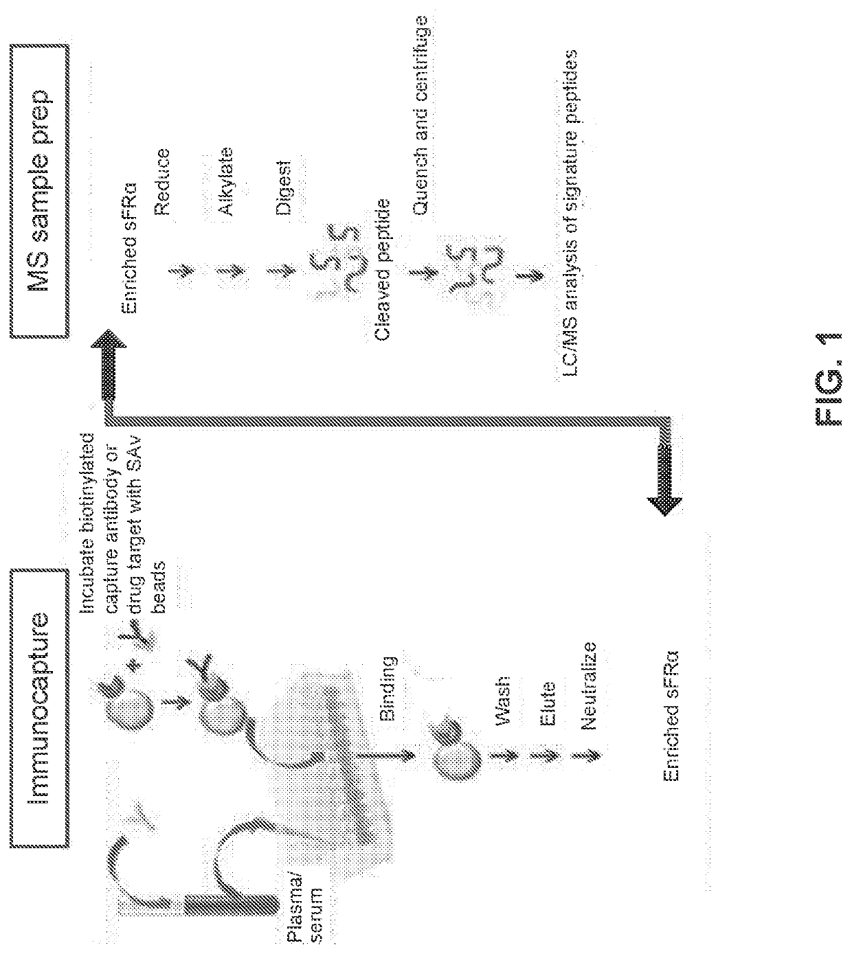
example 1
sFRα Immunocapture-LC / MS Assay
[0142]An ELISA-based assay was developed and described in WO 2014 / 036495. This assay was performed using ELISA plates coated with FOLR1-Fc (fusion peptide of huFOLR1 and murine IgG2A hinge, CH2, CH3), muFR1-9 as the capture antibody, and biotinylated muFR1-13. The assay was optimized by a systematic approach with the criteria being reproducible signals with a high signal to noise ratio, minimal matrix effects in human plasma samples, high repeatability and precision, and lowest limit of detection. The optimal ELISA conditions chosen resulted in detection of sFRα as low as 25 ng / mL with neglibible / low noise. Despite the optimized conditions, this ELISA assay did not provide enough sensitivity to distinguish between sFRα levels in normal subjects versus the range of distribution of sFRα levels in ovarian cancer patients to allow correlative analysis of sFRα levels and the disease state.
[0143]A more recent ELISA-based assay using two different human FRa-sp...
example 2
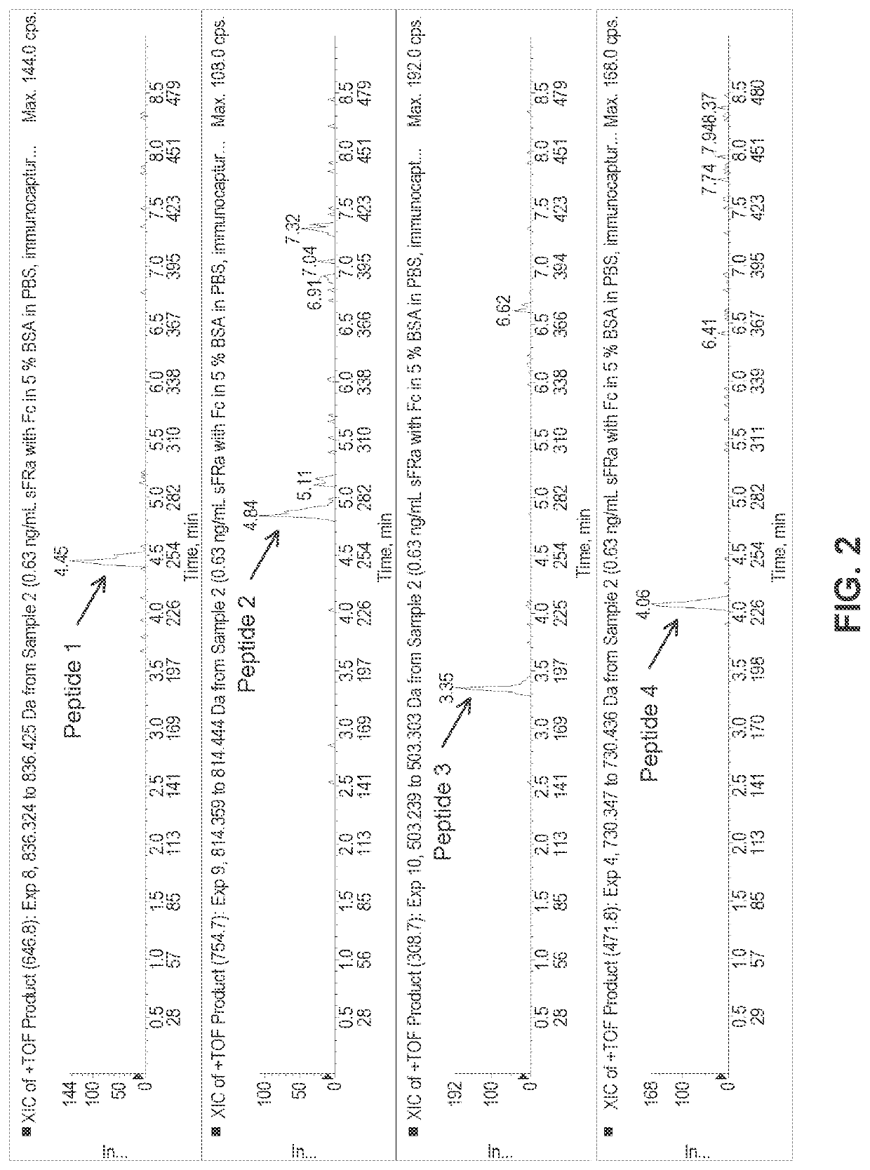
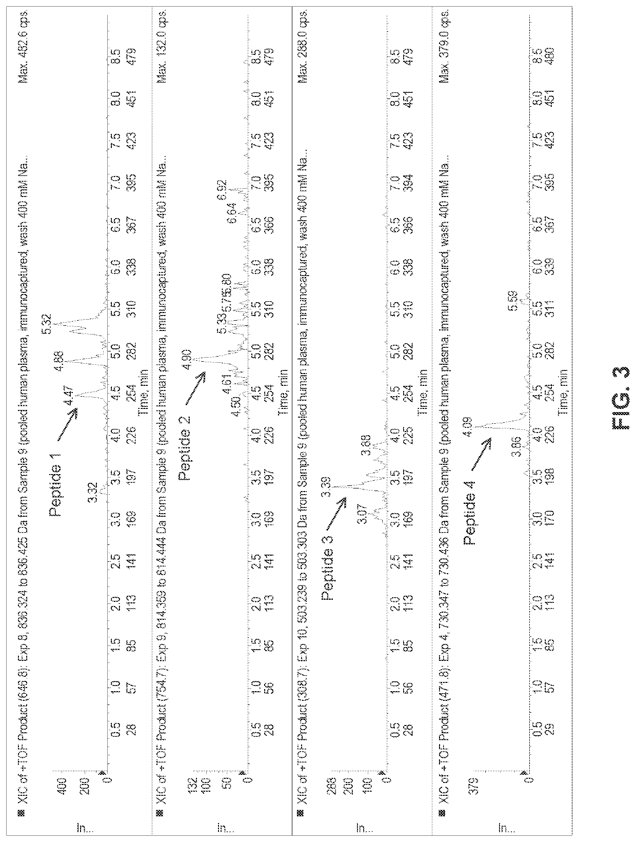
Data Analysis of Human Plasma Samples Using the sFRα Immunocapture-LC / MS Assay
[0148]Experiments were designed using the above parameters to measure the levels of sFRα in a sample containing low level of sFRα in surrogate matrix at 0.3 ng / mL. As shown in FIG. 2, the results of these experiments demonstrate that very low levels of sFRα can be measured by the immunocapture-LC / MS assay. Experiments were also designed to measure the levels of sFRα in a normal human plasma sample. As shown in FIG. 3, endogenous levels of sFRα, found around 0.5 ng / mL, can be measured by Immunocapture-LC / MS Assay. FIG. 4 shows an extracted ion chromatogram from a patient pre-dose sample with elevated sFRα. The results of this experiment demonstrates that patient plasma samples with elevated levels of sFRα can be clearly distinguished from that of endogenous levels of normal human plasma sample.
[0149]Experiments were designed to test the assay tolerability in the presence of a FRa-targeting immunoconjugate, ...
PUM
| Property | Measurement | Unit |
|---|---|---|
| pH | aaaaa | aaaaa |
| volume | aaaaa | aaaaa |
| liquid chromatography- | aaaaa | aaaaa |
Abstract
Description
Claims
Application Information