System and method for assessing a pulmonary image

Active Publication Date: 2020-10-08
KONINKLJIJKE PHILIPS NV
View PDF6 Cites 3 Cited by
  • Summary
  • Abstract
  • Description
  • Claims
  • Application Information

AI Technical Summary

Benefits of technology

The present invention provides a system, method, and computer program for assessing a pulmonary image to improve the detection of lung nodules in the image.

Problems solved by technology

However, determining the image quality just based on the noise is often not good enough, because the detectability of lung nodules in a pulmonary image might also depend on other factors.

Method used


Image

  • System and method for assessing a pulmonary image
  • System and method for assessing a pulmonary image
  • System and method for assessing a pulmonary image

Examples


Embodiment Construction

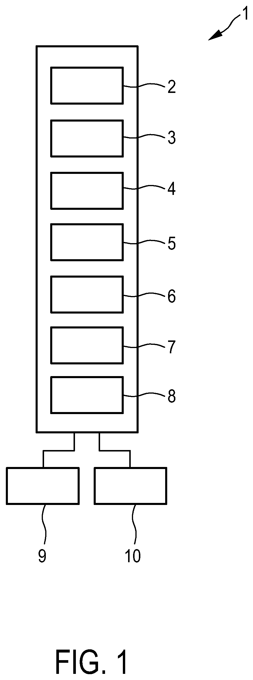
[0041]FIG. 1 shows schematically and exemplarily an embodiment of a system for assessing a pulmonary image. The system 1 comprises a pulmonary image providing unit 2 configured to provide a pulmonary image which comprises image elements to which image values are assigned and which shows lung vessels. In this embodiment the pulmonary image providing unit 2 is a storing unit in which the pulmonary image is stored, wherein the storing unit is adapted to provide the stored pulmonary image. Moreover, in this embodiment the pulmonary image is a computed tomography image of a lung of a patient. The patient is preferentially a human being. However, the patient can also be an animal.

[0042]The system 1 further comprises a smoothing unit 3 for spatially smoothing the provided pulmonary image with different degrees of smoothing, in order to generate different, smoothed pulmonary images. In this embodiment the smoothing unit 3 is adapted to apply a Gaussian image smoothing. The resulting pulmona...


PUM


Abstract

The invention relates to a system for assessing a pulmonary image which allows for an improved assessment with respect to lung nodules detectability. The pulmonary image is smoothed for providing different pulmonary images (20, 21, 22) with different degrees of smoothing, wherein signal values and noise values, which are indicative of the lung vessel detectability and the noise in these images, are determined and used for determining an image quality being indicative of the usability of the pulmonary image to be assessed for detecting lung nodules. Since a pulmonary image shows lung vessels with many different vessel sizes and with many different image values, which cover the respective ranges of potential lung nodules generally very well, the image quality determination based on the different pulmonary images with different degrees of smoothing allows for a reliable assessment of the pulmonary image's usability for detecting lung nodules. The image quality is used to determine a radiation dose level to be applied for generating a next pulmonary image.

Description

FIELD OF THE INVENTION[0001]The invention relates to a system, method and computer program for assessing a pulmonary image.BACKGROUND OF THE INVENTION[0002]For the detection of lung nodules pulmonary images like computed tomography images of the lung are often used, wherein for assessing the image quality of a pulmonary image with respect to its usability for reliably detecting a lung nodule its noise might be determined. However, determining the image quality just based on the noise is often not good enough, because the detectability of lung nodules in a pulmonary image might also depend on other factors.SUMMARY OF THE INVENTION[0003]It is an object of the present invention to provide a system, method and computer program for assessing a pulmonary image, which allows for an improved assessment with respect to the detectability of a lung nodule in the pulmonary image.[0004]In a first aspect of the present invention a system for assessing a pulmonary image is presented, wherein the s...

Claims


Application Information

Patent Timeline
IPC IPC(8): G06T7/00G06T7/11G06T5/00A61N5/10
CPCG06T2207/20021G06T7/11G06T2207/30064G06T2207/10081G06T7/0012A61N5/1031G06T2207/20182G06T5/002G06T2207/30168A61B6/032A61B6/50A61B6/504A61B6/5205A61B6/5258A61B6/542A61B6/545G06T2207/30061G06T5/70
Inventor WIEMKER, RAFAELNORDHOFF, TANJABUELOW, THOMASSAALBACH, AXELKLINDER, TOBIASBROSCH, TOMHARDER, TIM PHILIPPSOMMER, KARSTEN
Owner KONINKLJIJKE PHILIPS NV
Why Eureka
Try Eureka