System and Method to Quantify Tumor-Infiltrating Lymphocytes (TILs) for Clinical Pathology Analysis Based on Prediction, Spatial Analysis, Molecular Correlation, and Reconstruction of TIL Information Identified in Digitized Tissue Images

a tumor-infiltrating lymphocyte and clinical pathology technology, applied in image analysis, image enhancement, instruments, etc., can solve the problems of insufficient generating targeted and useful diagnostic assessments and/or refined classifications, set of input parameters that will not perform well across multiple images, and segmentation pipeline optimized for a tissue type will produce bad segmentation in images from other tissue types

Active Publication Date: 2020-12-10
THE RES FOUND TOR THE STATE UNIV OF NEW YORK +2
View PDF0 Cites 48 Cited by
  • Summary
  • Abstract
  • Description
  • Claims
  • Application Information

AI Technical Summary

Benefits of technology

[0027]In yet another disclosed embodiment, the computer readable device performs additional operations that include that any stain comprises hematoxylin and eosin (H&E) or Immunohostochemical stain. Yet other disclosed operations include that generating a refined TIL representation further comprises a single CNN predicting the TIL representations to generate a final TIL representation. Yet other disclosed operations include that the trained classification model is based on one or more of: a lymphocyte CNN and a necrosis CNN.

Problems solved by technology

Since there can be large variability in the color, texture, and morphology of tissues within and across cancer types (for example, heterogeneity can exist even within a tissue specimen such that the quality or state of the specimen manifests as non-uniform and / or diverse in character or content), it is likely that a set of input parameters will not perform well across multiple images.
It is not uncommon that a segmentation pipeline optimized for a tissue type will produce bad segmentations in images from other tissue types, and even in different regions of the same image.
However, human review of diagnostic tissue is qualitative, and is therefore proven to be prone to high amounts of inter-observer and / or intra-observer variability and hence, varied valuations rendering such review deficient in generating targeted and useful diagnostic assessments and / or refined classifications of cancer cells.
However, while TILs and spatial characterizations of TILs have shown significant value in diagnostic and prognostic settings, the ability to quantify TILs from diagnostic tissue has proven to be challenging, expensive, hard to scale, and is often subjective too.
The surge of digital pathology and impasse created from the proliferation of digitized whole slide diagnostic tissue images has necessarily resulted in datasets that are generally copious and burdensome to analyze in terms of differences and / or anomalies among such images of datasets.
Therefore, the useful analysis thereof and deduction of information such as quantification of useful values for classification and / or diagnosis of tumor cells has proven challenging.
Prior work in diagnostic imaging have either utilized multiple MC (ImmunoHistoChemical) stains or requires significant effort in tuning the algorithm.
Neither approach is generalizable or scalable, as the disclosed system and method and thus such prior methods are unlikely to be adopted in clinical care.

Method used


Image

  • System and Method to Quantify Tumor-Infiltrating Lymphocytes (TILs) for Clinical Pathology Analysis Based on Prediction, Spatial Analysis, Molecular Correlation, and Reconstruction of TIL Information Identified in Digitized Tissue Images
  • System and Method to Quantify Tumor-Infiltrating Lymphocytes (TILs) for Clinical Pathology Analysis Based on Prediction, Spatial Analysis, Molecular Correlation, and Reconstruction of TIL Information Identified in Digitized Tissue Images
  • System and Method to Quantify Tumor-Infiltrating Lymphocytes (TILs) for Clinical Pathology Analysis Based on Prediction, Spatial Analysis, Molecular Correlation, and Reconstruction of TIL Information Identified in Digitized Tissue Images

Examples


Embodiment Construction

[0078]In the following description, for the purposes of explanation, numerous specific details are set forth in order to provide a thorough understanding of example embodiments or aspects. It will be evident, however, to one skilled in the art, that an example embodiment may be practiced without all of the disclosed specific details.

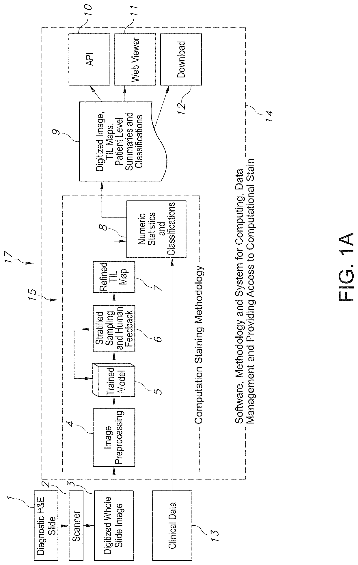
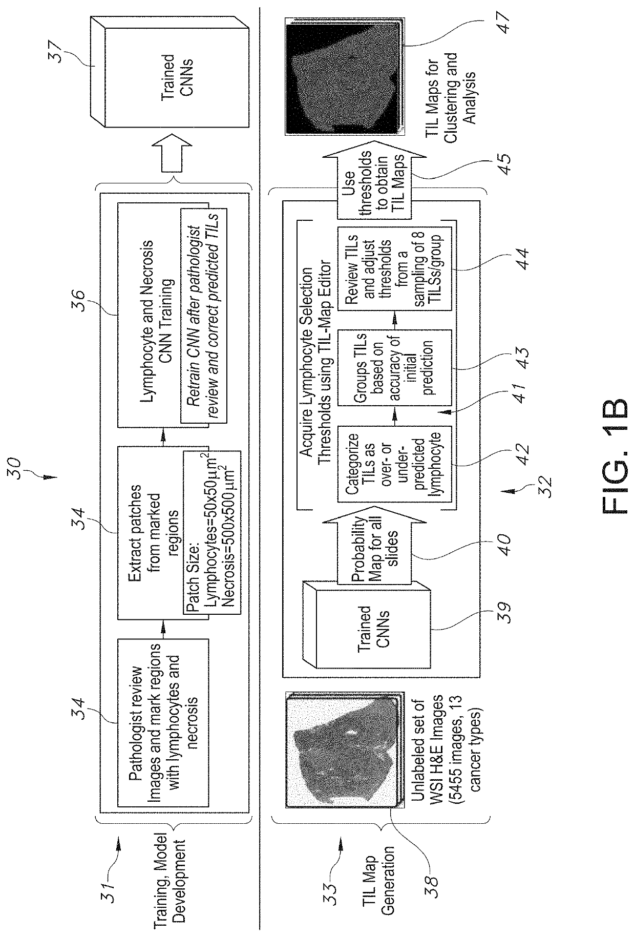
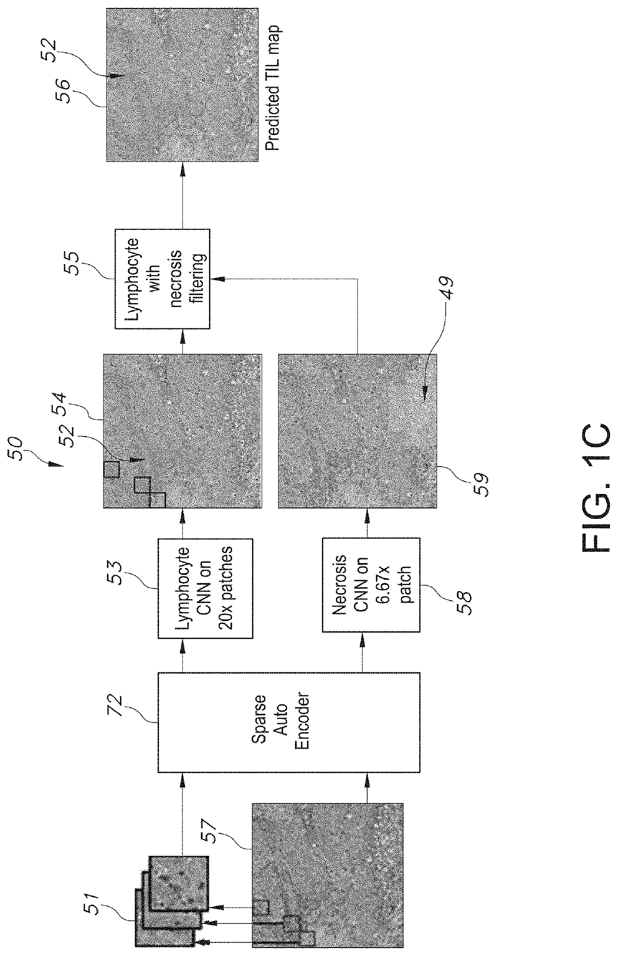
[0079]The present disclosure relates to a system and method associated with clinically processing, analyzing, and quantifying tumor-infiltrating lymphocytes (TILs) based on prediction, spatial analysis, molecular correlation, and reconstruction of TIL information associated with copious digital pathology images. Even more particularly, the present invention relates to a novel system and method that trains a classification model in order to predict the respective labeling of TILs associated with computationally stained and digitized whole slide images of Hematoxylin and Eosin (H&E) stained pathology specimens obtained from biopsied tissue, and spatially c...


PUM


Abstract

A system associated with quantifying a density level of tumor-infiltrating lymphocytes, based on prediction of reconstructed TIL information associated with tumoral tissue image data during pathology analysis of the tissue image data is disclosed. The system receives digitized diagnostic and stained whole-slide image data related to tissue of a particular type of tumoral data. Defined are regions of interest that represents a portion of, or a full image of the whole-slide image data. The image data is encoded into segmented data portions based on convolutional autoencoding of objects associated with the collection of image data. The density of tumor-infiltrating lymphocytes is determined of bounded segmented data portions for respective classification of the regions of interest. A classification label is assigned to the regions of interest. It is determined whether an assigned classification label is above a pre-determined threshold probability value of lymphocyte infiltrated. The threshold probability value is adjusted in order to re-assign the classification label to the regions of interest based on a varied sensitivity level of density of lymphocyte infiltrated. A trained classification model is generated based on the re-assigned classification labels to the regions of interest associated with segmented data portions using the adjusted threshold probability value. An unlabeled image data set is received to iteratively classify the segmented data portions based on a lymphocyte density level associated with portions of the unlabeled image data set, using the trained classification model. Tumor-infiltrating lymphocyte representations are generated based on prediction of TIL information associated with classified segmented data portions. A refined TIL representation based on prediction of the TIL representations is generated using the adjusted threshold probability value associated with the classified segmented data portions. A corresponding method and computer-readable device are also disclosed.

Description

CROSS-REFERENCE TO RELATED APPLICATION(S)[0001]The present application is the U.S. National Phase of, and claims priority of International Patent Application No. PCT / US2018 / 063231, filed on Nov. 30, 2018, which claims the benefit of both U.S. Provisional Application No. 62 / 592,931, filed on Nov. 30, 2017, and U.S. Provisional Application No. 62 / 651,677 filed on Apr. 2, 2018, the specifications of which are each incorporated by reference herein, in their entirety for all purposes.STATEMENT OF GOVERNMENT LICENSE RIGHTS[0002]This invention was made with government support under CA180924, CA143835, CA199461, and HHSN261200800001E awarded by the National Institutes of Health. The government has certain rights in the invention.FIELD OF THE DISCLOSURE[0003]The present disclosure relates to a system and method associated with clinically processing, analyzing, and analyzing tumor-infiltrating lymphocytes (TILs) based on prediction, spatial analysis, molecular correlation, and reconstruction ...

Claims


Application Information

Patent Timeline
Patent Type & Authority Applications(United States)
IPC IPC(8): G06T7/00G06K9/32G06T9/00G06K9/62G06T7/11G06V10/25G06V10/764G06V10/774
CPCG06K9/3233G06T2207/30096G06T2207/20084G06T7/0012G06T2207/20076G06T7/11G06K9/6256G06T2207/20081G06T9/00G06K9/6267G06T7/143G06T2207/10056G06T2207/30024G06T2207/10024G06V20/695G06V20/698G06V10/25G06V10/267G06V10/454G06V10/82G06V10/764G06V10/774G06F18/2413G06F18/24G06F18/214
Inventor SALTZ, JOEL HASKINKURC, TAHSINGUPTA, RAJARSIZHAO, TIANHAOBATISTE, REBECCAHOU, LENGUYEN, VUSAMARAS, DIMITRIOSRAO, ARVINDVAN ARNAM, JOHNSINGH, PANKAJLAZAR, ALEXANDERSHARMA, ASHISHSHMULEVICH, ILYATHORSSON, VESTEINN
Owner THE RES FOUND TOR THE STATE UNIV OF NEW YORK
Why Eureka
Try Eureka