

High-resolution spatial macromolecule abundance assessment
a macromolecule and spatial technology, applied in the field of spatial assessment of macromolecule abundance, can solve the problems of low degree of multiplexing with a high degree of technical difficulty, and/or provide only low resolution of spatial capture across an array
- Summary
- Abstract
- Description
- Claims
- Application Information
AI Technical Summary
Benefits of technology
Problems solved by technology
Method used
Image

Examples
example 1
and Methods
Beads:
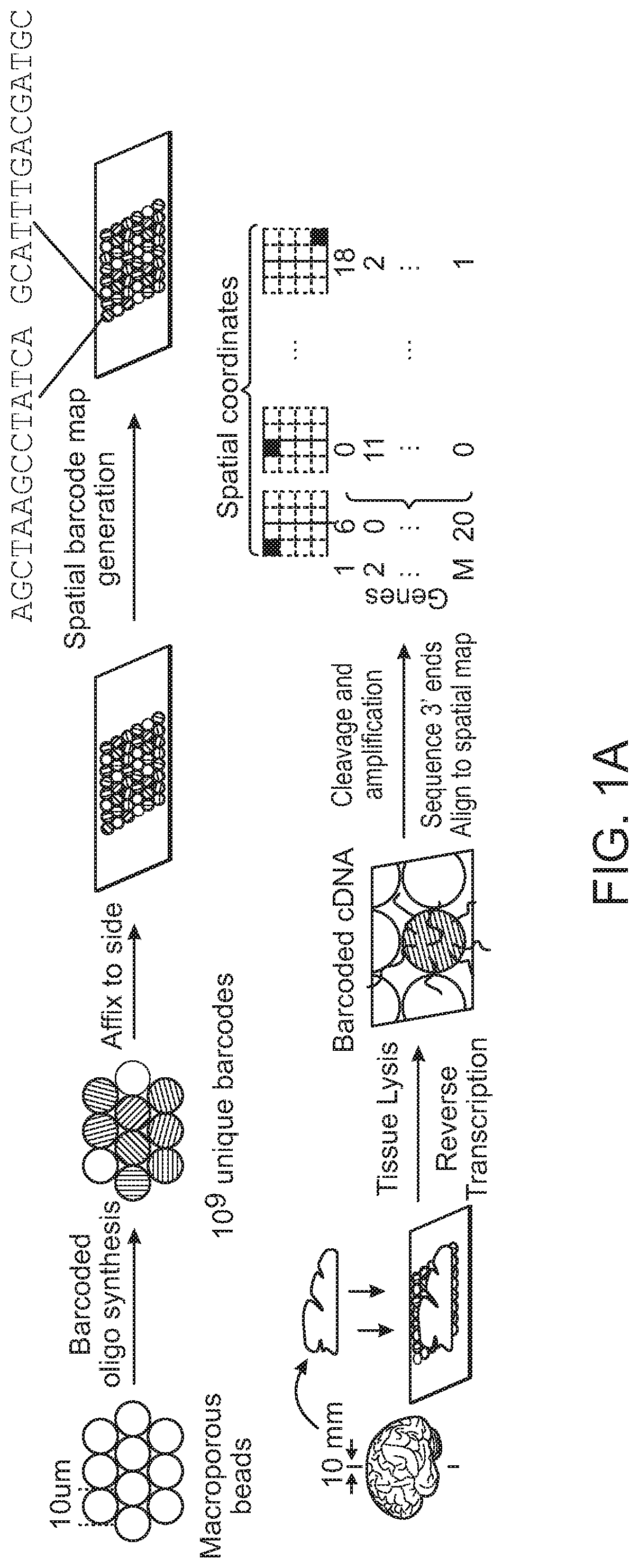
[0212]Beads were produced by the ChemGenes Corporation on one of two polystyrene supports (Agilent and Custom Polystyrene supports from AM Biotech). Beads were used with one of the two following sequences:
Sequence 1:(SEQ ID NO: 1)5'-PEG Linker-TTTT-PCT-GCCGGTAATACGACTCACTATAGGGCTACACGACGCTCTTCCGATCTJJJJJJTCTTCAGCGTTCCCGAGAJJJJJJJNNNNNNNNT30 Sequence 2:(SEQ ID NO: 2)5'-Linker-TTTTTTTTGCCGGGGCTACACGACGCTCTTCCGATCTJJJJJJJJTCTTCAGCGTTCCCGAGAJJJJJJJNNNNNNNNT30
Here, PCT represents a photocleavable thymidine; J bases represent bases generated by split-pool barcoding, such that every oligo on a given bead has the same J bases; Ns represent bases generated by mixing, so every oligo on a given bead has different N bases; and T30 represents a string of 30 thymidines. The two sequences corresponded to different bead batches, which were not found to differ significantly in terms of the number of transcripts per bead.
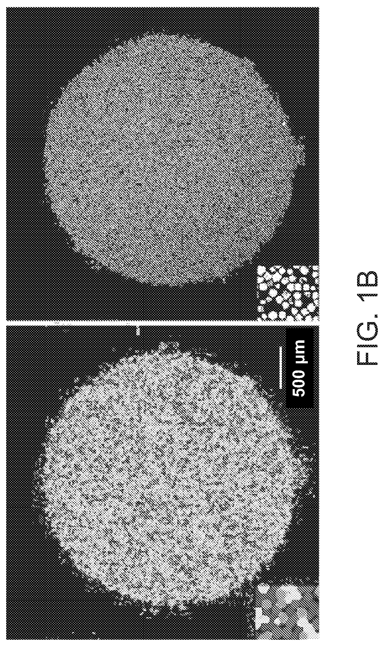
Puck Preparation:
[0213]Pucks were prepared as follows. Glass c...
example 2
sociation of Individually Barcode-Tagged Microbeads with a Glass Slide Provided a High-Resolution Array for Transcriptome Capture
[0286]A large number of 10 μm beads that possessed unique nucleic acid barcodes were prepared via methods as described previously (e.g., as set forth in WO 2016 / 040476). Specifically, to generate a population of beads possessing individual barcodes that could be used for identification of an individual bead's position when arranged in a two-dimensional array as presently exemplified, polynucleotide synthesis was performed upon the surface of the beads in a pool-and-split fashion such that in each cycle of synthesis the beads were split into subsets that were subjected to different chemical reactions; and then this split-pool process was repeated in multiple cycles, to produce a combinatorially large number (approaching 4n) of distinct nucleic acid barcodes (FIG. 1A). Nucleotides were chemically built onto the bead material in a high-throughput manner, and ...
example 3
lide-Associated Barcode-Tagged Microbead Array Captured Transcriptomes with Robust Spatial Resolution
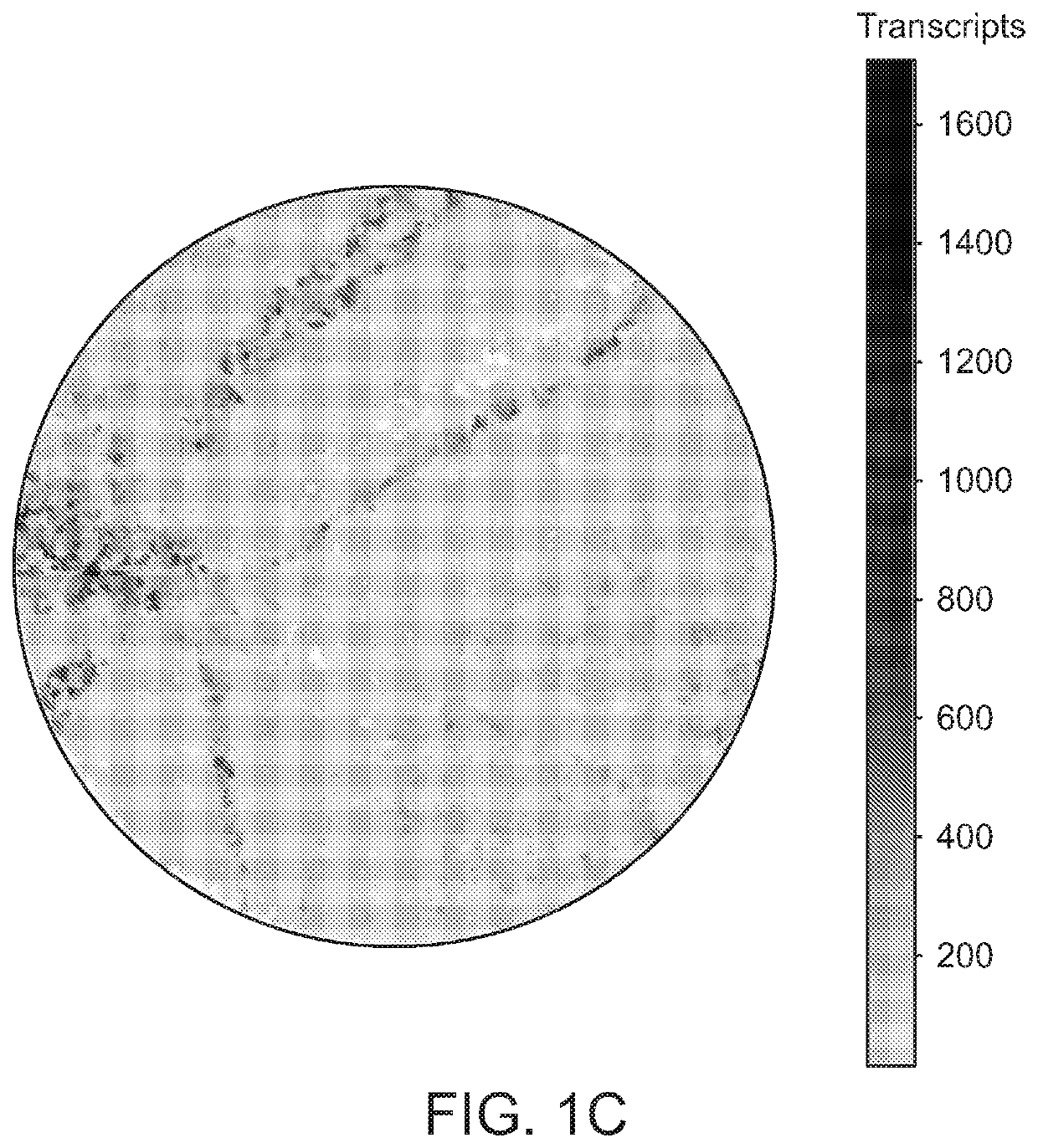
[0290]To determine if the surface could capture RNA with high resolution, a protocol was developed wherein frozen tissue sections (˜10 μm) were transferred onto the bead surface via cryosectioning (7). This process efficiently transferred RNA from the tissue to the surface, and subsequent processing of beads via standard single-cell library preparation pipelines generated 3′-end digital expression libraries. Performing this process on mouse hippocampal tissue slices, the distribution of transcripts across the puck was found to have recapitulated the distribution of cell bodies observed in the tissue (FIG. 1C). By comparing the width of CA1 observed in Slide-seq hippocampal data to that width observed in an adjacent, DAPI-stained tissue section (FIG. 1D), it was estimated that the length-scale of lateral diffusion of transcripts during hybridization was less than the width of an indiv...
PUM
| Property | Measurement | Unit |
|---|---|---|
| Temperature | aaaaa | aaaaa |
| Temperature | aaaaa | aaaaa |
| Temperature | aaaaa | aaaaa |
Abstract
Description
Claims
Application Information