Eureka AIR delivers breakthrough ideas for toughest innovation challenges, trusted by R&D personnel around the world.

Spectrally encoded optical polarization imaging for detecting skin cancer margins

a technology of optical polarization imaging and skin cancer, applied in the field of methods and systems for imaging tumors, can solve the problems of increasing the number of stages needed, reducing the accuracy of the detection method, so as to improve the treatment effect, save time, and use resources effectively

Pending Publication Date: 2021-08-19
UNIV OF MASSACHUSETTS
View PDF2 Cites 0 Cited by
  • Summary
  • Abstract
  • Description
  • Claims
  • Application Information

AI Technical Summary

Benefits of technology

This patent is about a new technology that uses advanced imaging to improve the accuracy of surgical margins for skin cancer. The technology is fast, easy to use, and produces clear images that can be easily understood without extensive training. Compared to other imaging techniques, optical polarization imaging (OPI) is the preferred method because it is harmless to human skin and can provide adequate imaging depth and resolution. The patent also mentions that other technologies can also be used with the subject technology. Overall, the patent describes how to improve the surgical margin assessment process for skin cancer and similar lesions by utilizing advanced imaging strategies.

Problems solved by technology

However, determining the initial dimensions of the first excision stage prior to surgery is challenging and relies on the surgeon's clinical intuition and experience.
The inaccurate, initial identification of the tumor margins increases the number of stages needed to clear the tumor, amplifying the cost of and time required for the surgical procedure.
While RCM has a very high resolution and can identify individual tumor cells, RCM has small field of view and produces images that are challenging to acquire and interpret without extensive training.
OCT can assess larger areas of skin than RCM, but has a lower lateral resolution and cannot discriminate individual tumors cell reliably.
Both technologies are comparatively slow and expensive.
Additionally, not all skin cancers are operable.

Method used


Image

  • Spectrally encoded optical polarization imaging for detecting skin cancer margins
  • Spectrally encoded optical polarization imaging for detecting skin cancer margins
  • Spectrally encoded optical polarization imaging for detecting skin cancer margins

Examples


Embodiment Construction

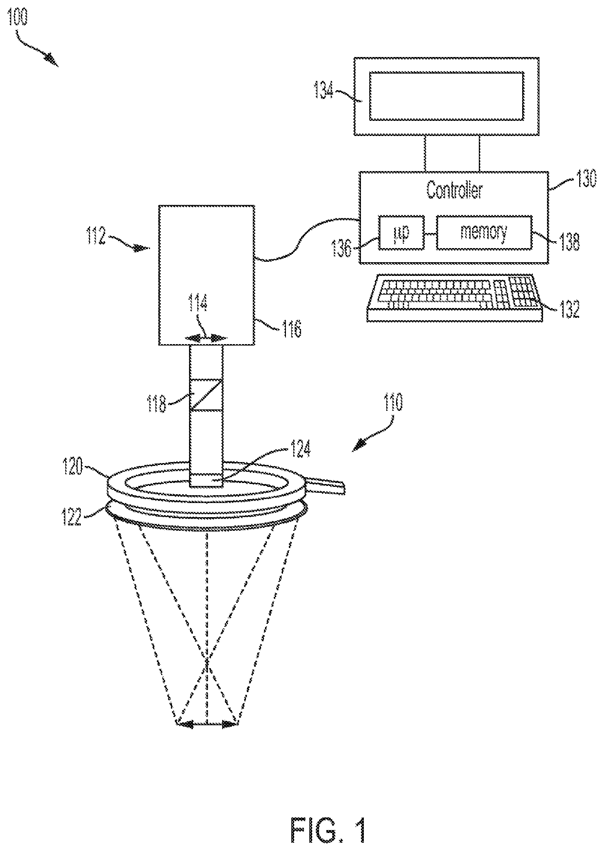
[0018]The subject technology overcomes many of the prior art problems associated with accurately determining margins of skin cancer such as for preoperative delineation of keratinocytic carcinoma (KC) boundaries or pre- and post-treatment of inoperable skin cancer. The advantages, and other features of the systems and methods disclosed herein, will become more readily apparent to those having ordinary skill in the art from the following detailed description of certain preferred embodiments taken in conjunction with the drawings which set forth representative embodiments of the present invention and wherein like reference numerals identify similar structural elements. It is understood that references to the figures such as up, down, upward, downward, left, and right are with respect to the figures and not meant in a limiting sense.

[0019]In brief overview, the subject optical polarization imaging (OPI) technology can be used for the determination of the margins of any skin lesion or c...


PUM


Abstract

Many treatments of skin cancer need an accurate assessment of the margins of the tumor. Spectrally-encoded optical polarization imaging improves upon the demarcation of skin cancer such as keratinocytic carcinomas. When the clinician can outline the clinical boundary of each lesion, surgery and other treatments can be more efficient and effective. Optical images of the lesions are acquired at various wavelengths, then spectrally-encoded. Spectral-encoding of the images minimizes the impact of background pigmentation and vascularization so that the tumor can be accurately visualized.

Description

BACKGROUND1. Field[0001]The subject disclosure relates to methods and systems for imaging tumors for improved determination of tumor margins.2. Background of the Related Art[0002]Keratinocytic carcinoma (KC) is the most common form of cancer in the United States, diagnosed in 3.3 million individuals annually. Surgical removal of the cancerous lesion is the most common form of treatment. Mohs micrographic surgery has been the standard for KC tumor removal for several decades. Intraoperative histologic identification and removal of tumor at the surgical margin yields very high cure rates and helps to preserve normal tissue compared to other techniques.[0003]However, determining the initial dimensions of the first excision stage prior to surgery is challenging and relies on the surgeon's clinical intuition and experience. Even the most experienced surgeons underestimate or miscalculate the clinical margins 35% of the time on average. The inaccurate, initial identification of the tumor ...

Claims


Application Information

Patent Timeline
IPC IPC(8): A61B5/00
CPCA61B5/444A61B5/0075A61B5/742A61B5/7203A61B5/0077A61B2576/02A61B5/7217G16H30/40
Inventor YAROSLAVSKY, ANNA N.
Owner UNIV OF MASSACHUSETTS
Why Eureka
Eureka Blog
Learn More