
Chromosomal enhancement and automatic detection of chromosomal abnormalities using chromosomal ideograms

a technology of chromosomal ideograms and enhancement, which is applied in the field of chromosomal enhancement and automatic detection of chromosomal abnormalities using chromosomal ideograms, can solve the problems of difficult detection and often missed chromosomal abnormalities, and achieve accurate and sensitive abnormalities identification and easy analysis

Pending Publication Date: 2021-09-23
NAT GUARD HEALTH AFFAIRS +2
View PDF0 Cites 3 Cited by
  • Summary
  • Abstract
  • Description
  • Claims
  • Application Information

AI Technical Summary

Benefits of technology

The invention provides a way to detect abnormalities in chromosomal images using a method and system that converts micrographic images into an intermediate phase. This system can accurately and sensitively identify chromosomal abnormalities, which could help in the diagnosis and treatment of genetic disorders.

Problems solved by technology

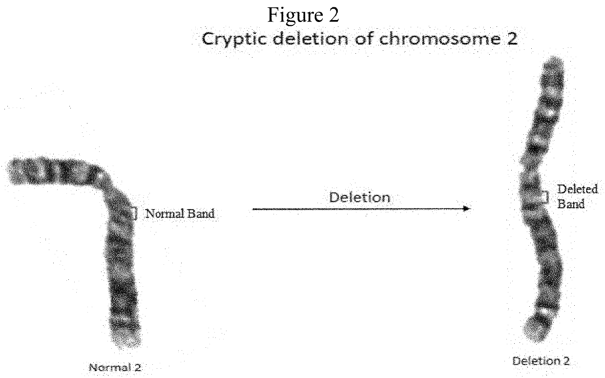
However, certain chromosomal abnormalities, such as short, cryptic deletions to a chromosome (FIG. 2A) are difficult to detect and often require review by a cytogeneticist with years of experience.
Thus, chromosomal abnormalities often get missed because of the complex efforts required to analyze micrographic images of chromosomes containing abnormalities.

Method used


Image

  • Chromosomal enhancement and automatic detection of chromosomal abnormalities using chromosomal ideograms
  • Chromosomal enhancement and automatic detection of chromosomal abnormalities using chromosomal ideograms
  • Chromosomal enhancement and automatic detection of chromosomal abnormalities using chromosomal ideograms

Examples


examples

[0118]The following graphic examples of certain embodiments of the system or methods disclosed herein are described by FIGS. 18 (Process 1), 19 (Process 2) and 20 (Process 3).

[0119]In Process 1, an image of stained chromosomes is produced (first panel), which is further clarified using conventional enhancement, such as decreasing background and increasing contrast (panel 2). Panel 3 shows the subsequent generation of a karyogram, which in panel 4 are separated into pairs of single chromosomal images. Process 1 describes a conventional process of producing a karyogram.

[0120]Process 2 describes inputting chromosomal images into a trained Cycle-GAN model, which outputs ideograms; or when given an ideogram, outputs chromosomal images. This process which is disclosed herein facilitates the identification of chromosomal abnormalities that are undetectable or difficult to accurately detect by conventional methods. The output of process 2, option 2, is used as a single chromosome.

[0121]Proc...


PUM


Abstract

A method for detecting chromosomal abnormalities comprising conversion of a chromosomal image into an ideogram which is compared to a control or standard ideogram of a chromosome; or converting an ideogram of a chromosome into a chromosomal image.

Description

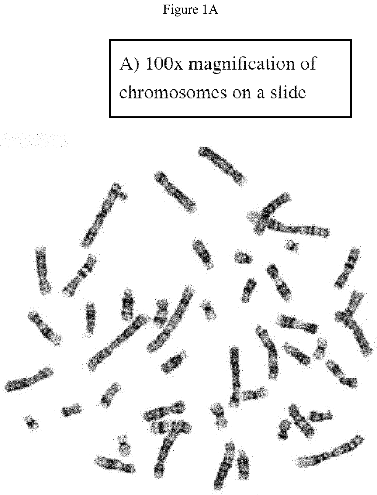
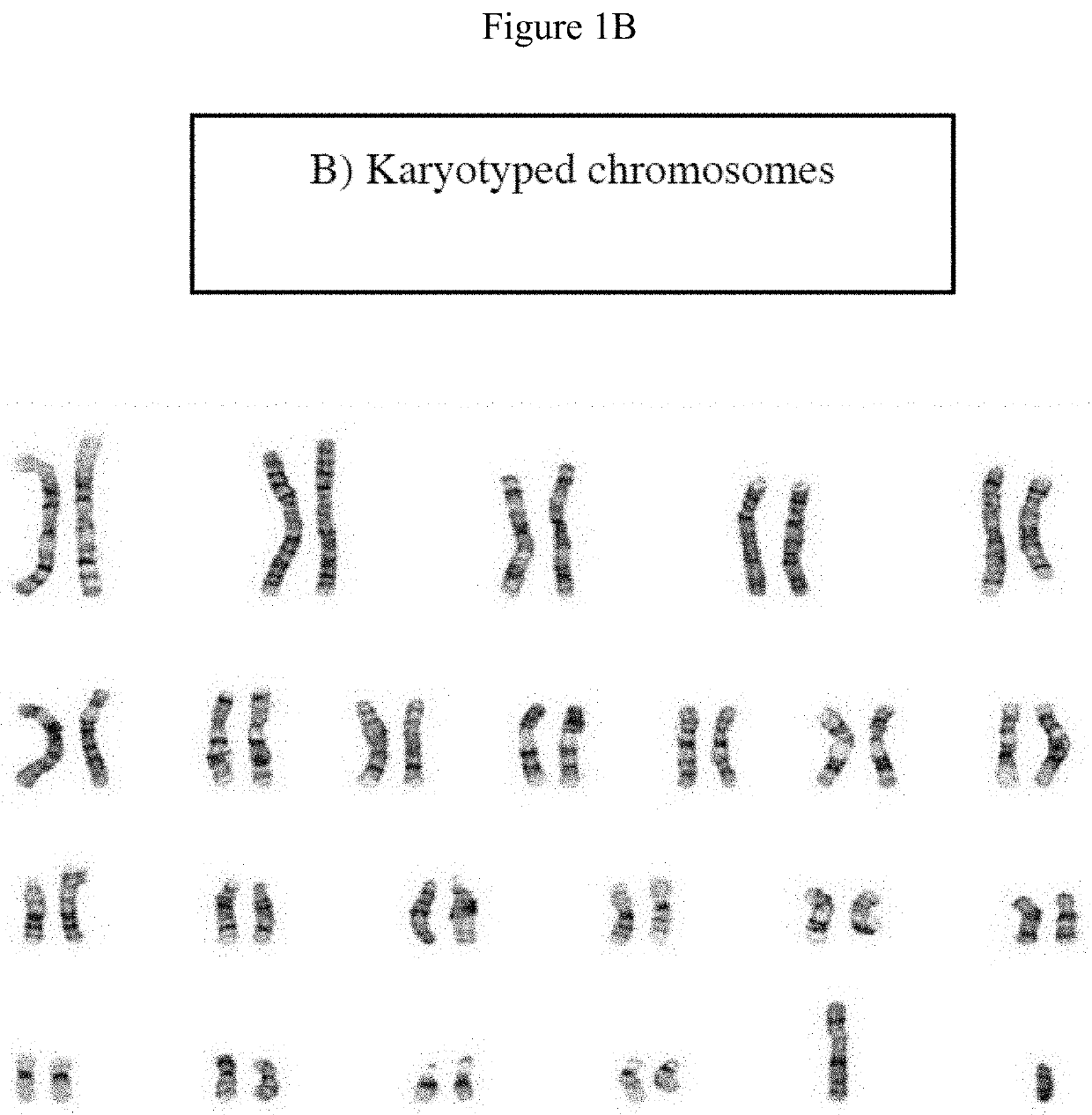
CROSS-REFERENCE TO RELATED APPLICATION[0001]This application claims priority to U.S. Provisional Application No. 62 / 990,713, filed Mar. 17, 2020, which is hereby incorporated by reference for all purposes.BACKGROUND OF THE INVENTIONField of Invention[0002]Cytogenetic medical instrumentation and methods using artificial intelligence.Description of Related Art[0003]Chromosomal analysis or karyotyping is typically performed in a cytogenetics laboratory. Karyotyping is a process in which the number and visual appearance of the chromosomes in the cell nuclei of an organism or species is determined. Existing protocols use a microscope which can visualize metaphase chromosomes (FIG. 1A), a camera to photograph the chromosomes, and software to help classify and organize chromosomes in a sample (FIG. 1B).[0004]Conventional procedures can detect abnormalities in chromosome number, for example, a missing or additional chromosome, or large abnormalities such as deletion of a portion of a chromo...

Claims


Application Information

Patent Timeline
Patent Type & Authority Applications(United States)
IPC IPC(8): G06T7/00G16B45/00G16B20/20G06T5/00
CPCG06T7/0014G16B45/00G06T2207/30072G06T5/006G06T2207/20081G16B20/20G16B40/20G06T2207/10056G06T2207/20084G06T5/77G06T5/70G06T5/60G06T5/80
Inventor BOKHARI, YAHYA ABDULFATTAHALHAREERI, AREEJ ABDULAZIZALKHALDI, AZIZAH MOHAMMEDALJOUIE, ABDULRHMAN FAHAD
Owner NAT GUARD HEALTH AFFAIRS
Learn More