
Image processing apparatus, medical imaging apparatus, and image processing program

a technology of image processing and medical imaging, applied in image enhancement, healthcare informatics, image analysis, etc., can solve the problems of affecting the image quality, so as to reduce the noise of the image with different noise levels, reduce the cost of calculation, and improve the effect of image quality

Pending Publication Date: 2021-12-30
HITACHI LTD
View PDF0 Cites 0 Cited by
  • Summary
  • Abstract
  • Description
  • Claims
  • Application Information

AI Technical Summary

Benefits of technology

The invention allows for the removal of noise from an image that has varying levels of noise in different parts of the image. This is done quickly and at a low cost. The resulting image is then improved to meet the preferences of the reader.

Problems solved by technology

In a medical imaging apparatus such as a magnetic resonance imaging (hereinafter, referred to as MRI) apparatus, an X-ray computed tomography (CT) apparatus, and an ultrasonic diagnostic apparatus, if it takes a long time for capturing in order to acquire data (or a signal) for reconstructing an image, an adverse effect such as a burden on a subject or appearance of an image artifact due to movement of the subject is exerted.
However, in the high-speed capturing method of the MRI apparatus, since an amount of data used for the image is smaller than usual in order to increase the speed, noise is generated and image quality is deteriorated.
In addition, a noise distribution in the same capturing plane is not uniform with respect to a capturing space.
Further, in an ultrasonic capturing apparatus, noise is particularly increased in a deep region or the like where sensitivity is insufficient.
For this reason, a calculation cost for filter generation increases.
In addition, since the generated nonlinear noise filter is sequentially applied to the images, the calculation cost also increases at the time of filter processing.
Further, since the image viewed by a reader is an image after the image quality is improved by the generated filter, there is a problem that a preference of the reader cannot be reflected in adjustment of the filter itself.

Method used

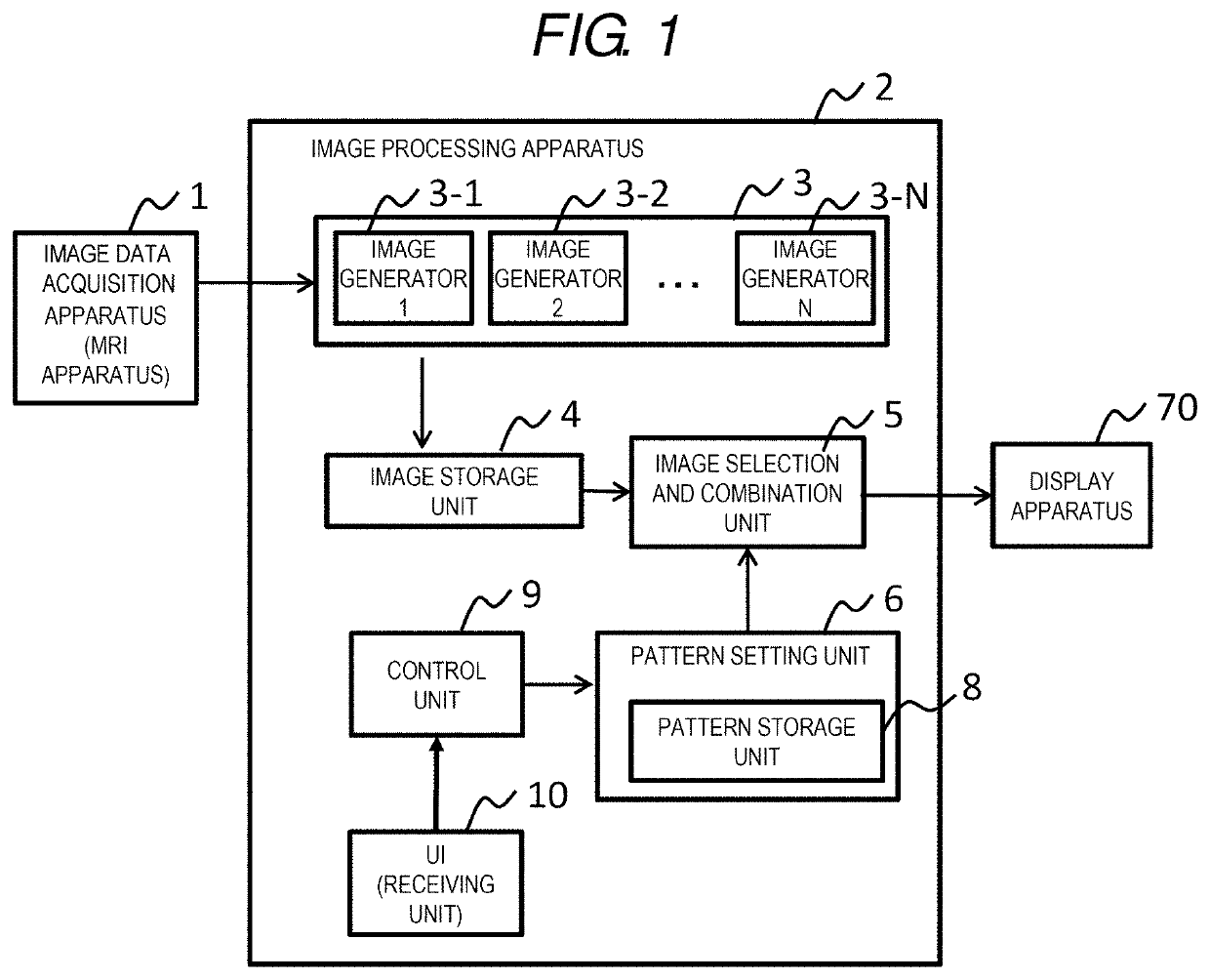

Image

  • Image processing apparatus, medical imaging apparatus, and image processing program
  • Image processing apparatus, medical imaging apparatus, and image processing program
  • Image processing apparatus, medical imaging apparatus, and image processing program

Examples


first embodiment

[0021]A medical imaging apparatus according to a first embodiment includes an MRI apparatus as an image data acquisition apparatus. The medical imaging apparatus according to the first embodiment will be described with reference to FIGS. 1 to 6.

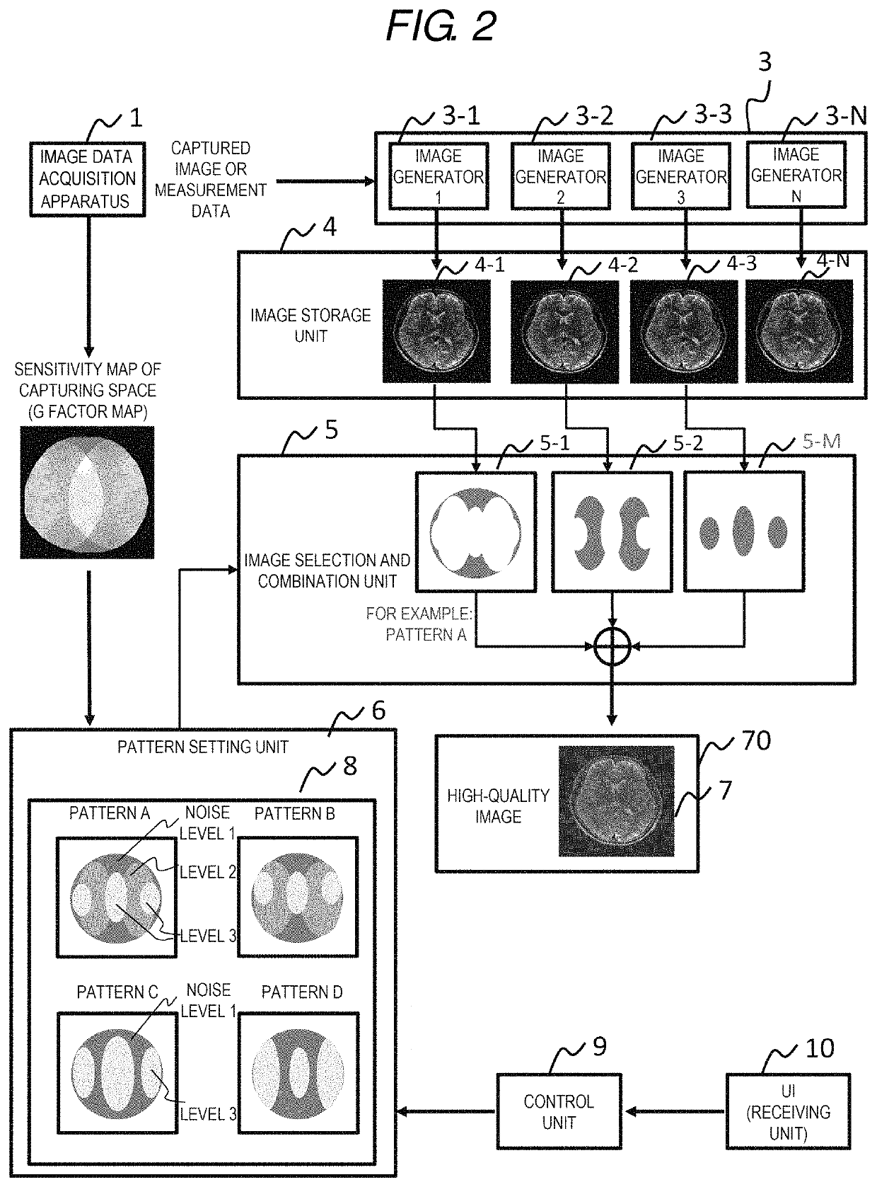
[0022]As shown in FIGS. 1 and 2, the medical imaging apparatus according to the first embodiment includes an image data acquisition apparatus 1 and an image processing apparatus 2. The image data acquisition apparatus 1 is the MRI apparatus.

2>

[0023]The image processing apparatus 2 includes a plurality of image generators 3-1 to 3-N, an image selection and combination unit 5, an image storage unit 4, a pattern setting unit 6, a receiving unit 10, and a control unit 9.

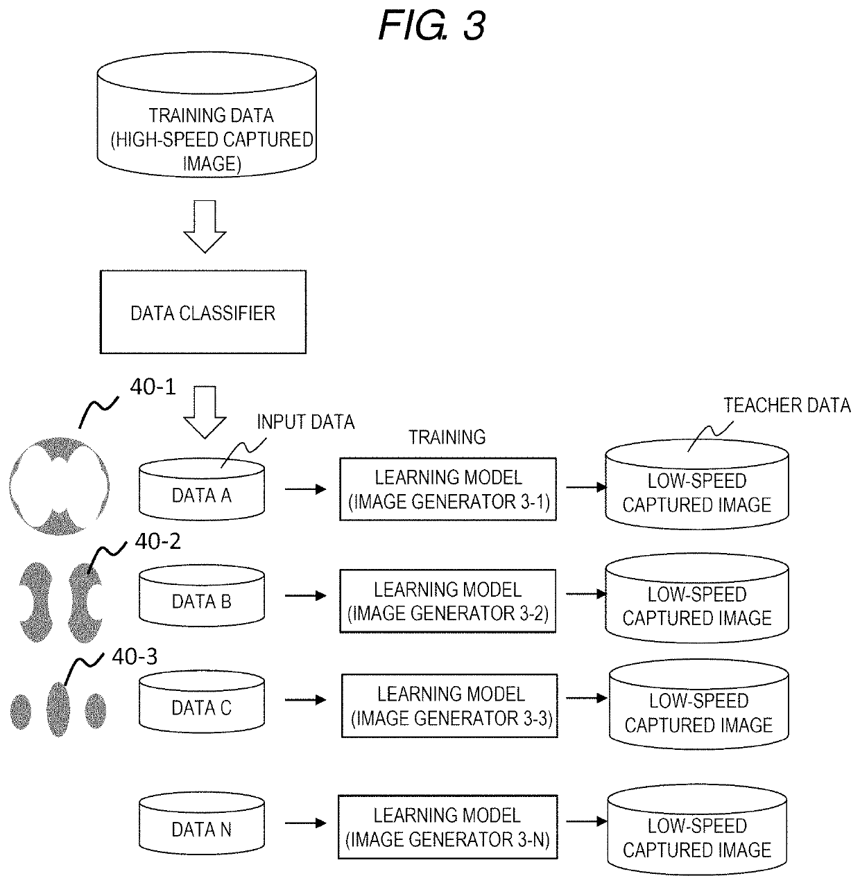
[0024]The image generators 3-1 to 3-N receive measurement data or a captured image (referred to as an original image) obtained by the image data acquisition apparatus 1, and generate different images 4-1 to 4-N for a same imaging range as the original image. Specifically, the imag...

second embodiment

[0064]As a medical imaging apparatus according to a second embodiment, an ultrasonic imaging apparatus is provided as the image data acquisition apparatus 1. The medical imaging apparatus according to the second embodiment will be described with reference to FIGS. 7 to 9.

2>

[0065]As shown in FIGS. 7 and 8, the configuration of the image processing apparatus 2 is similar to that of the image processing apparatus 2 according to the first embodiment. Since a noise level of an ultrasonic image increases as a depth of a subject increases, a region selection pattern is a pattern in which a region having a shallowest noise level 1 is selected and a region which is deeper as the noise level increases is selected.

[0066]The image processing apparatus 2 according to the second embodiment includes an imaging condition reception and pattern selection unit 11. The imaging condition reception and pattern selection unit 11 receives the imaging condition from the image data acquisition apparatus 1, s...


PUM


Abstract

Provided are an image processing apparatus, a medical imaging apparatus, and an image processing program that remove noise from an image having different noise levels depending on regions in the image at a low calculation cost, and enable high quality according to a preference of a reader. A plurality of image generators receive measurement data or a captured image obtained by an image data acquisition apparatus and generate different images for a same imaging range. An image selection and combination unit selects different image regions from a plurality of images generated by the plurality of image generators according to a predetermined region selection pattern, and generates one image by combining the images of the selected image regions.

Description

CROSS-REFERENCE TO RELATED APPLICATION[0001]The present application claims priority from Japanese application JP2020-109420, filed on Jun. 25, 2020, the contents of which is hereby incorporated by reference into this application.BACKGROUND OF THE INVENTION1. Field of the Invention[0002]The present invention relates to an apparatus that generates a medical image with reduced noise.2. Description of the Related Art[0003]In a medical imaging apparatus such as a magnetic resonance imaging (hereinafter, referred to as MRI) apparatus, an X-ray computed tomography (CT) apparatus, and an ultrasonic diagnostic apparatus, if it takes a long time for capturing in order to acquire data (or a signal) for reconstructing an image, an adverse effect such as a burden on a subject or appearance of an image artifact due to movement of the subject is exerted. Therefore, a high-speed capturing method for shortening a capturing time by devising a capturing method is developed in each modality.[0004]For e...

Claims


Application Information

Patent Timeline
IPC IPC(8): G06T5/00G16H30/40G16H30/20G06T7/00
CPCG06T5/002G06T7/0012G16H30/20G16H30/40G06F3/04845G06F3/04847G06F3/04855G06F3/0482G06T2207/10088G06T2207/20081G06T2207/20084G06T5/70G06T2207/20221G06T5/50G06T5/60
Inventor TANAKA, CHIZUEKANEKO, YUKIO
Owner HITACHI LTD
Learn More