Harmonic shear wave imaging

a shear wave and imaging technology, applied in the field of tissue shear wave elastography, can solve the problems of inability to achieve the optimal clinical use of a separate driver, the transducer type, and the ultrasonic frequency to produce biases in the shear wave speed measurement,

Pending Publication Date: 2022-01-27
PENN STATE RES FOUND
View PDF1 Cites 0 Cited by
  • Summary
  • Abstract
  • Description
  • Claims
  • Application Information

AI Technical Summary

Benefits of technology

[0006]Push pulses are interleaved with imaging / detection pulses to measure and reconstruct maps of shear wave speed. The imaging pulses may be plane-wave, which allow measuring displacements in a region of the tissue of interest. However, other types of detection pulses can also be used. Applying imaging / detection pulses between push pulses allows generating and measuring shear wave propagation simultaneously.
[0014]According to the embodiments of the present invention, narrowband low frequency excitation and visualization of shear waves are made possible.

Problems solved by technology

A drawback of transient methods is that factors such as ultrasound frequencies, transducer types, and imaging depths produce biases in the shear wave speed measurements.
However, the need of a separate driver may not be optimal for clinical use because the added complexity to the measurement.

Method used


Image

  • Harmonic shear wave imaging
  • Harmonic shear wave imaging
  • Harmonic shear wave imaging

Examples


Embodiment Construction

Overview of the Method

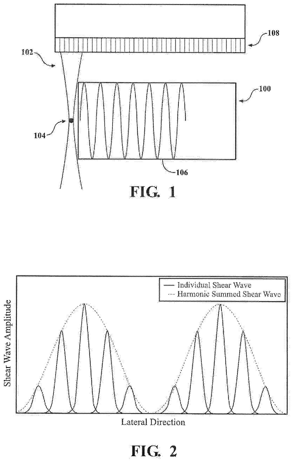
[0028]Changes in the stiffness of soft tissues are often related to the presence of pathological conditions. Ultrasound elastography imaging techniques provide noninvasive ways to measure tissue stiffness. In dynamic ultrasound elastography, tissue stiffness is quantitatively evaluated by inducing shear waves in the tissue and monitoring the speed of the shear wave propagation, based on the fact that the shear wave speed is positively proportional to tissue stiffness.

[0029]The present invention provides embodiments of a shear wave ultrasound elastography method that allows measurements that may be more robust, and / or more accurate, more consistent.

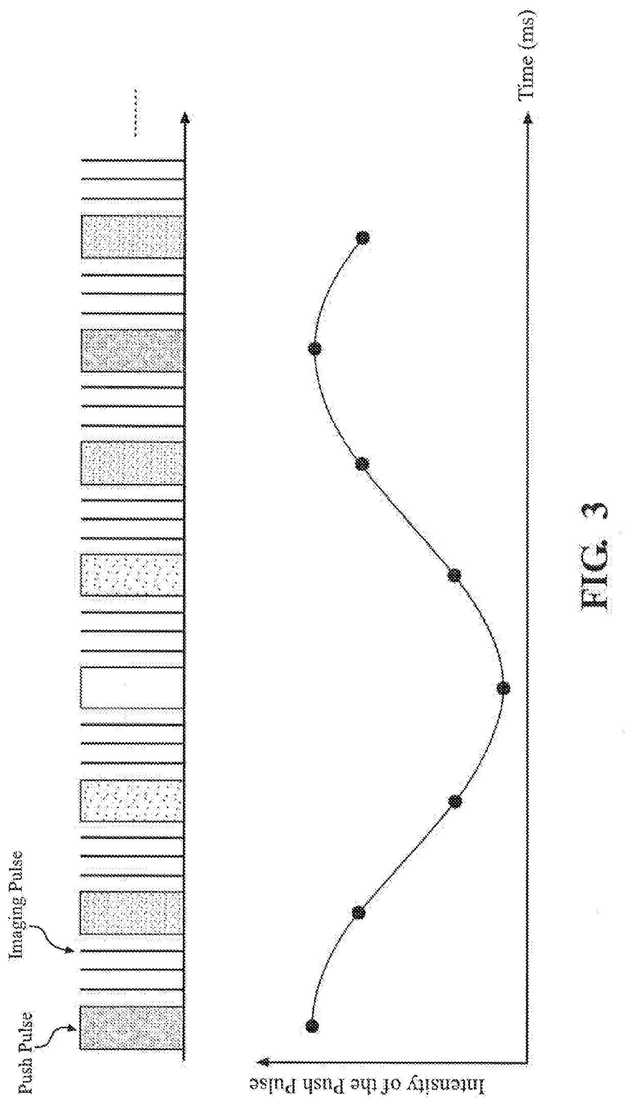
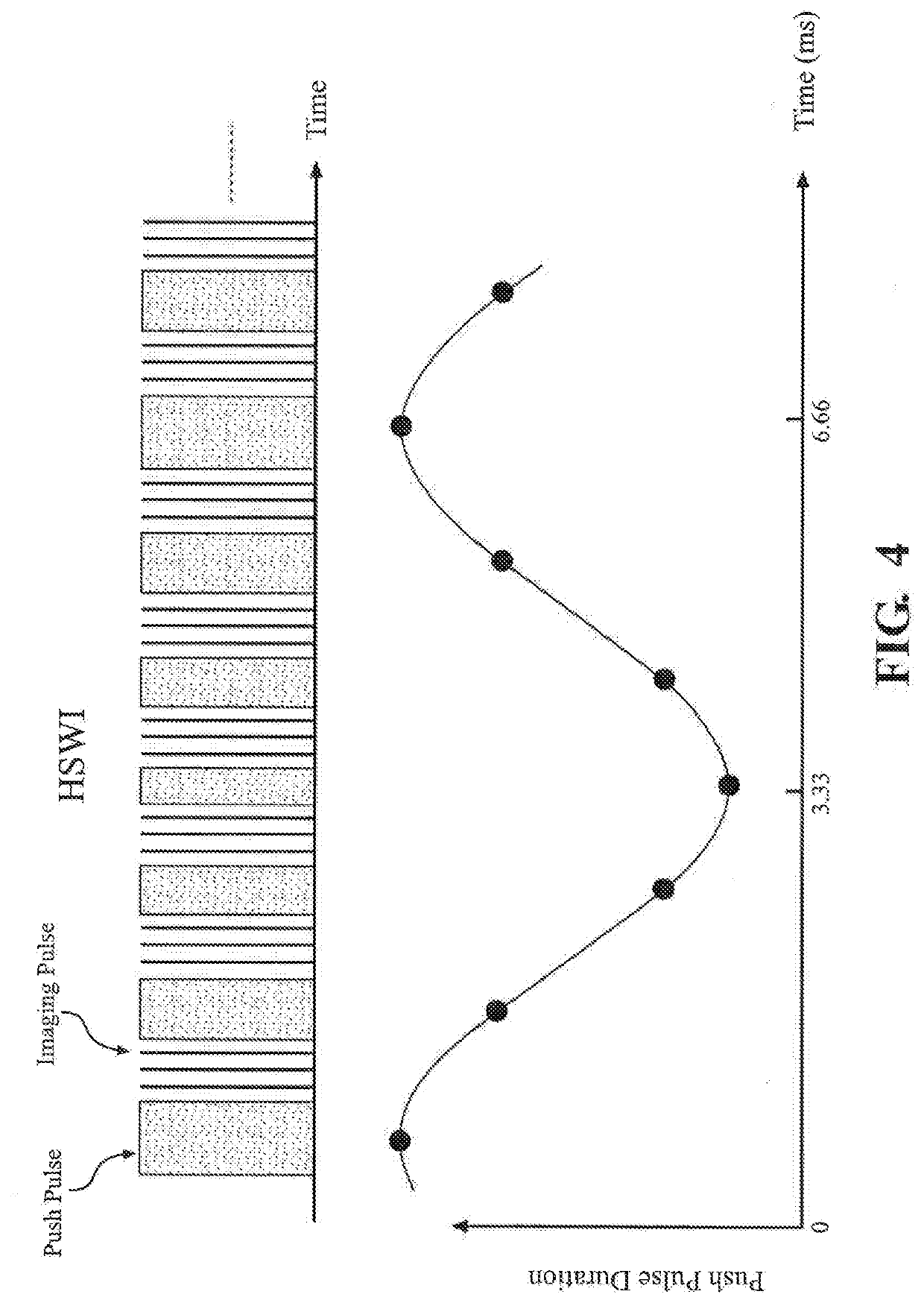
[0030]An embodiment of the present invention provides a shear wave generation method. The shear wave is generated by transmitting periodically-modulated ultrasound “push” pulses with a primary modulation frequency using a transducer. The method, also referred to as harmonic shear wave imaging ((HSWI), successively tra...


PUM


Abstract

A method of performing shear wave elastography in tissue includes transmitting successively a series of ultrasound push pulses in the tissue in a region of interest (ROI) using a single array transducer. The acoustic intensities of the push pulses are sinusoidally modulated with a modulation frequency, Each push pulse generates an acoustic radiation force that pushes the tissue and creates an individual shear wave propagating through the tissue. The amplitudes of the shear waves, and therefore, the displacements produced by the push pulses, are positively proportionally to the intensities of the push pulses. The successively created individual shear waves with different amplitudes sum together to form a continuous, harmonic summed shear wave with a single frequency the same as the modulation frequency of the push pulses.

Description

CROSS REFERENCE TO RELATED APPLICATION[0001]This application is the U.S. National Stage of PCT / US2019 / 062911 filed Nov. 25, 2019, which claims priority to U.S. Provisional Patent Application Ser. No. 62 / 772,913, filed Nov. 29, 2018, the entire content of both are incorporated herein by reference in their entirety.FIELD OF THE INVENTION[0002]The present invention relates to shear wave elastography in tissues.BACKGROUND OF THE INVENTION[0003]The shear modulus of soft tissues has important clinical implications since it often changes as a consequence of disease or injury. Therefore, the shear modulus can be a sensitive biomarker for diagnosing and monitoring a variety of clinical conditions. Elastography is a generic name given to methods for measuring the shear modulus non-invasively in the body. Elastography requires the generation of shear wave in the tissue of interest and measurement and analysis of the shear wave speed using an imaging method or other noninvasive techniques. The ...

Claims


Application Information

Patent Timeline
Patent Type & Authority Applications(United States)
IPC IPC(8): A61B8/08G01S7/52
CPCA61B8/485A61B8/483G01S7/52022G01S7/52042G01S15/8915
Inventor CORREALES, DANIEL HUMBERTO CORTESLIN, CHE-YUSADEGHI, SEYEDALI
Owner PENN STATE RES FOUND
Why Eureka
Try Eureka