Method and Probe System for Tissue Analysis in a Surgical Cavity in an Intraoperative Procedure

a tissue analysis and intraoperative procedure technology, applied in the field of tissue analysis in the surgical cavity in an intraoperative procedure, can solve the problems of limited area coverage, high probability of cancerous tissue remaining in the patient, and inability to provide the assessment of the entire cavity

Pending Publication Date: 2022-03-17
CYTOVERIS INC
View PDF0 Cites 0 Cited by
  • Summary
  • Abstract
  • Description
  • Claims
  • Application Information

AI Technical Summary

Benefits of technology

[0024]According to another aspect of the present disclosure, a tissue cavity probe is provided that includes a stem, a probe body, an optical scanning assembly, and at least one light conduit. The probe body is connected to the stem. The probe body has at least one optically transparent wall that defines an interior cavity of the probe body. The optical scanning assembly is disposed within the interior cavity of the probe body. The at least one light conduit is configured to be connected to one or more light sources and in communication with the optical scanning assembly. The optical scanning assembly is configured to direct one or more beams of excitation light to be incident with a tissue defining at least part of a tissue cavity, and the probe is configured to collect at least one response from the tissue defining the at least part of the tissue cavity and transfer the at least one response to the at least one light conduit.
[0025]In any of the aspects or embodiments described above and herein, the optical scanning assembly may be controllable to direct the one or more excitation light beams to be incident with a plurality of different regions of the tissue defining the at least part of the tissue cavity.
[0026]The foregoing features and elements may be combined in various combinations without exclusivity, unless expressly indicated otherwise. These features and elements as well as the operation thereof will become more apparent in light of the following description and the accompanying drawings. It should be understood, however, the following description and drawings are intended to be exemplary in nature and non-limiting.

Problems solved by technology

This often results in what are referred to as positive margins of the excised specimen; i.e., indications of cancerous tissue either on, or in close proximity to, the surface of the excised specimen, meaning there this a very high likelihood of cancerous tissue remaining in the patient.
However, they are limited in area coverage and do not provide the assessment of the entire cavity.

Method used


Image

  • Method and Probe System for Tissue Analysis in a Surgical Cavity in an Intraoperative Procedure
  • Method and Probe System for Tissue Analysis in a Surgical Cavity in an Intraoperative Procedure
  • Method and Probe System for Tissue Analysis in a Surgical Cavity in an Intraoperative Procedure

Examples


Embodiment Construction

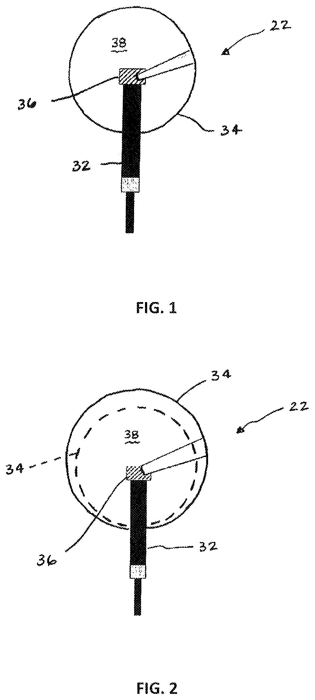
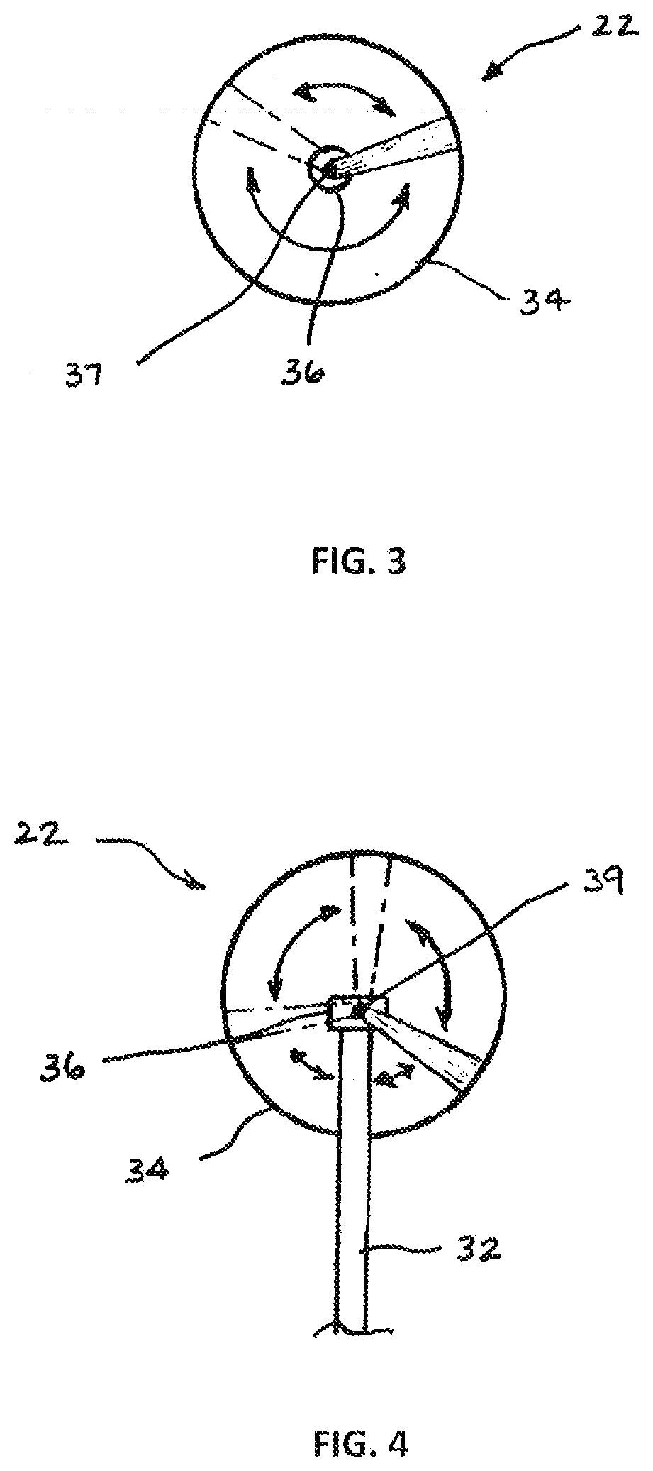
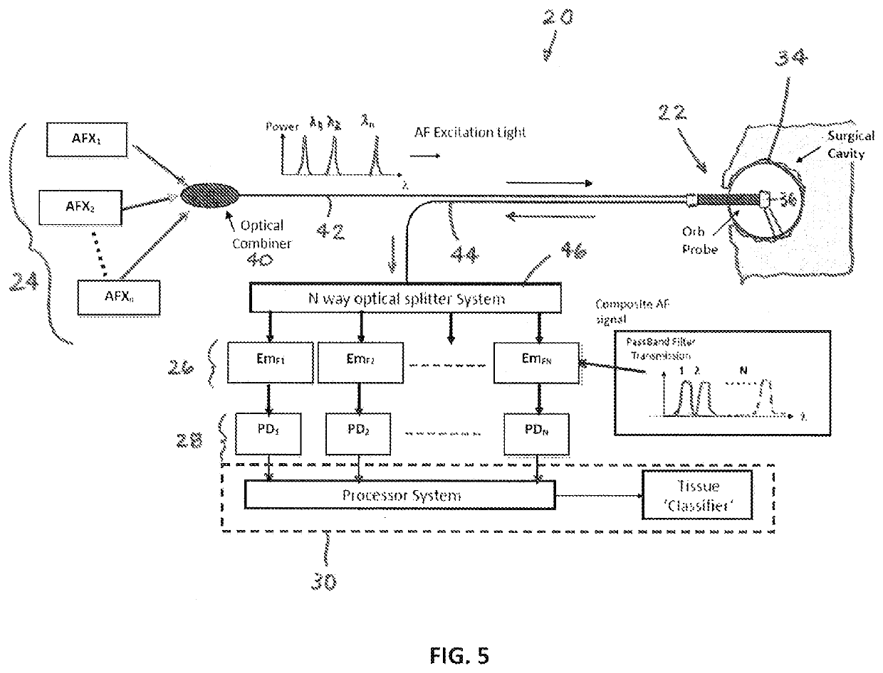
[0034]Aspects of the present disclosure include a novel and unobvious system 20 that utilizes one or more optical spectroscopic techniques such as Raman spectroscopy, tissue autofluorescence (AF), or diffuse reflectance, or near-infrared (NIR) absorption, or the like to assess the status of tissue defining a tissue cavity for the presence of diseased tissue (e.g., cancerous tissue) following a resection procedure. Diffuse reflectance refers to reflected excitation light that may be detected in a diminished intensity due to absorption and / or scattering of the interrogating excitation light within the tissue. To facilitate the description herein, the present disclosure will be described as utilizing Raman spectroscopy and AF. The present disclosure is not, however, limited to Raman, AF or combination thereof. The present disclosure is applicable to almost any form of tissue resection surgery and provides particular utility for cancer tissue resection surgery.

[0035]The system 20 includ...


PUM


Abstract

A system and method for determining the presence of cancerous tissue within a tissue cavity is provided. The system includes one or more excitation light sources, a photodetector, a probe, and a system controller. The probe includes an optically transparent probe body configured to fit within the tissue cavity. The system controller is in communication with the excitation light sources, the photodetector, and a memory storing instructions. The instructions when executed cause the system controller to a) control the excitation light sources to produce excitation light beams within the probe body, the excitation light beams operable to produce a response of the tissue to the interrogation and control the photodetector to detect the response and produce signals representative thereof; and b) produce information indicative of a presence of the cancerous tissue using the signals representative of the response.

Description

[0001]This application claims priority to U.S. Patent Appln. No. 63 / 079,738 filed Sep. 17, 2020, which is hereby incorporated by reference in its entirety.BACKGROUND OF THE INVENTION1. Technical Field[0002]The present disclosure relates to devices and methods for assessing diseased tissue in general, and the devices and methods for assessing residual diseased tissue in an intraoperative procedure in particular.2. Background Information[0003]For many decades the reference method for the diagnosis of cancer has been histopathological examination of tissues using conventional microscopy. This process is known as surgical pathology. In surgical pathology, tissue samples can be produced from surgical procedures (tumor resection), diagnostic biopsies or autopsies. These tissue samples are subsequently subjected to a process that includes dissection, fixation, and cutting of tissue into precisely thin slices which are stained for contrast and mounted onto glass slides. The slides are exami...

Claims


Application Information

Patent Timeline
Patent Type & Authority Applications(United States)
IPC IPC(8): A61B5/00
CPCA61B5/0091A61B5/0071A61B5/0075
Inventor FOURNIER, DAVIDPANDEY, RISHIKESHSAPACK, MICHAEL A.CURRY, PATRICK M.KERSEY, ALAN
Owner CYTOVERIS INC
Why Eureka
Try Eureka