Ultrasound imaging system and method for generating an enhanced image

Active Publication Date: 2022-03-24
GE PRECISION HEALTHCARE
View PDF10 Cites 0 Cited by
  • Summary
  • Abstract
  • Description
  • Claims
  • Application Information

AI Technical Summary

Benefits of technology

The patent aims to fix the issues mentioned earlier in the text. The technical effects sought by the patent include improvements to overcome the difficulties and difficulties faced by the existing technology.

Problems solved by technology

One problem with using higher frequencies or predominantly higher frequencies to generate ultrasound images is that while higher frequencies of ultrasound have higher resolution, they do not penetrate as well as lower frequencies.
A shadow region in an image may make it difficult or impossible for a clinician to accurately understand the patient's anatomy and / or make proper measurements based on an image using conventional ultrasound imaging techniques.

Method used


Image

  • Ultrasound imaging system and method for generating an enhanced image
  • Ultrasound imaging system and method for generating an enhanced image
  • Ultrasound imaging system and method for generating an enhanced image

Examples


first embodiment

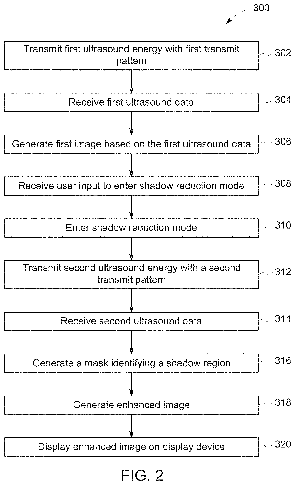
[0046]At step 316, the processor 116 generates a mask based on either the first ultrasound data or the second ultrasound data. the processor 116 may generate the mask based on the first ultrasound data. For example, the processor 116 may generate the mask based on the first ultrasound data after it has been beamformed by the receive beamformer 110, but before it has been scan-converted for display on the display device 118. For purposes of this disclosure, we may refer to ultrasound data as an “image” after it has been beamformed. According to other embodiments, the processor 116 may generate the mask based on the first ultrasound data after it has been scan-converted and is ready for display as an image on the display device 118.

second embodiment

[0047] the processor 116 may generate the mask based on the second ultrasound data. The processor 116 may generate the mask based on the second ultrasound data after it has been beamformed by the receive beamformer 110, but before it has been scan-converted for display on the display device 118. According to other embodiments, the processor 116 may generate the mask based on the second ultrasound data after it has been scan-converted and is ready for display as an image on the display device 118.

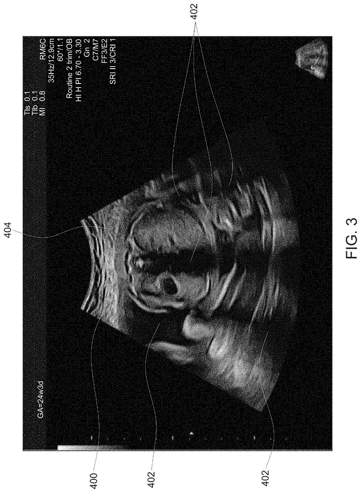
[0048]The processor 116 is configured to identify a shadow region based on either the first ultrasound data or the second ultrasound data. The shadow region may be a single connected region or a plurality of separate shadow subregions. The shadow region is the portion of an ultrasound image where the data lacks enough intensity to generate a diagnostically useful image. FIG. 3 shows an image 400 generated based on ultrasound data. In the example shown in FIG. 3, the image 400 has been scan-c...


PUM


Abstract

A method and ultrasound imaging system includes transmitting first ultrasound energy with a first transmit pattern, receiving first ultrasound data based on the first ultrasound energy, and generating a first image based on the first ultrasound data. The method and system includes entering a shadow reduction mode and performing the following steps in the shadow reduction mode: transmitting second ultrasound energy with a second transmit pattern configured to have greater penetration than the first transmit pattern, receiving second ultrasound data based on the second ultrasound energy, generating a mask identifying a shadow region and generating an enhanced image including an enhanced shadow region, wherein the second ultrasound data is used to generate the enhanced shadow region, and displaying the enhanced image.

Description

FIELD OF THE INVENTION[0001]This disclosure relates generally to an ultrasound imaging system and a method for generating and displaying an enhanced image including an enhanced shadow region.BACKGROUND OF THE INVENTION[0002]Ultrasound imaging is an imaging modality based on echoes received from transmitted ultrasound energy. Ultrasound imaging has many clinical uses including general imaging, cardiac imaging, musculoskeletal imaging (MSK), point-of-care, primary care, and obstetrics / women's health. Ultrasound imaging has many advantages as an imaging modality. The ultrasound energy used to acquire images is non-ionizing and therefore safer for many applications than other imaging modalities that rely on X-ray or other ionizing forms of radiation. Additionally, ultrasound imaging is a relatively affordable imaging modality, which makes it well-suited for widespread use in a variety of clinical scenarios.[0003]In ultrasound imaging, image quality is typically related to a signal-to-no...

Claims


Application Information

Patent Timeline
IPC IPC(8): A61B8/08A61B8/00
CPCA61B8/5207A61B8/481G01S15/8979A61B8/54A61B8/42A61B8/44A61B8/463A61B8/4444A61B8/4488A61B8/465A61B8/486A61B8/488A61B8/483A61B8/466A61B8/5246A61B8/5269G01S7/52046G01S15/8952G01S15/8959A61B8/5223G16H30/40G16H50/20G06T7/0012G06T2207/10132G06T2207/20084
Inventor FOSODEDER, ERWINSCHWARZ, GERALDSWEET, CAROLYN ELIZABETH
Owner GE PRECISION HEALTHCARE
Why Eureka
Try Eureka