Image guided high intensity focused ultrasound device for therapy in obstetrics and gynecology

a high-intensity, ultrasound technology, applied in the field of real-time ultrasound imaging, can solve the problems of reducing the power of the hifu transferred to the tissue, etc., and achieve the effect of enhancing the propagation of the hifu beam and negatively affecting the hifu treatmen

a high-intensity, ultrasound technology, applied in the field of real-time ultrasound imaging, can solve the problems of reducing the power of the hifu transferred to the tissue, etc., and achieve the effect of enhancing the propagation of the hifu beam and negatively affecting the hifu treatmen

US20050203399A1Inactive Publication Date: 2005-09-15UNIV OF WASHINGTON

Image

  • Image guided high intensity focused ultrasound device for therapy in obstetrics and gynecology
  • Image guided high intensity focused ultrasound device for therapy in obstetrics and gynecology
  • Image guided high intensity focused ultrasound device for therapy in obstetrics and gynecology

Examples


Embodiment Construction

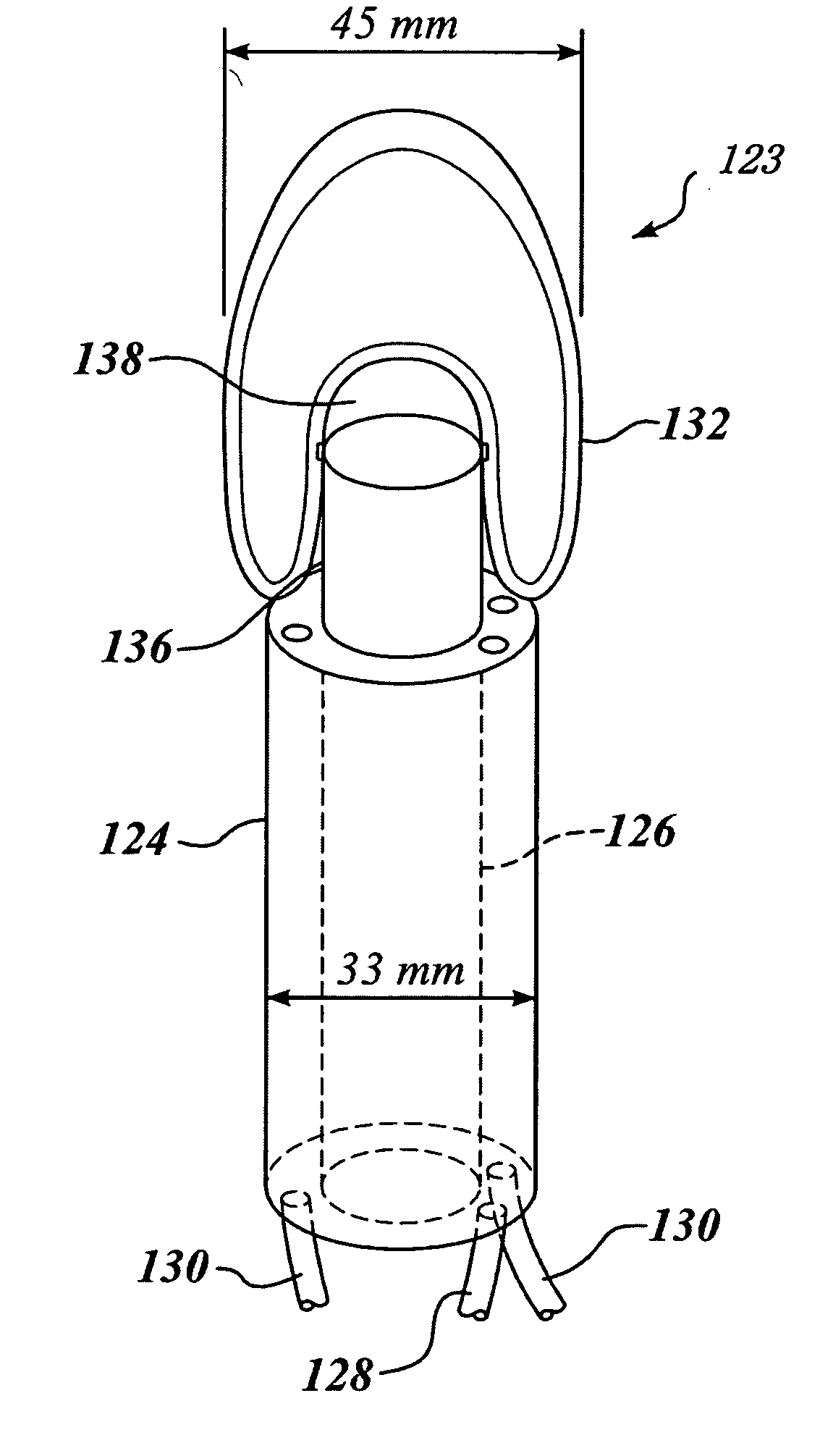
[0053] The terms “therapeutic transducer,”“HIFU transducer,” and “high intensity transducer,” as used herein and in the claims that follow all refer to a transducer that is capable of being energized to produce ultrasonic waves that are much more energetic than the ultrasonic pulses produced by an imaging transducer, and which can be focused or directed onto a discrete location, such as a treatment site in a target area. However, in at least one embodiment of the present invention, not all ultrasonic waves produced by such a transducer are necessarily at a high intensity, as is explained below.

[0054] When administering HIFU therapy, it is very desirable to be able to observe a treatment site, to ensure that lesions induced by the HIFU therapy are being produced at the desired location. Failure to properly aim the HIFU beam will result in undesired tissue necrosis of non target tissue. From a practical standpoint, this goal has not proven easy to accomplish when ultrasound is used t...


PUM


Abstract

A frame ensures that the alignment between a high intensity focused ultrasound (HIFU) transducer designed for vaginal use and a commercially available ultrasound image probe is maintained, so that the HIFU focus remains in the image plane during HIFU therapy. A water-filled membrane placed between the HIFU transducer and the treatment site provides acoustic coupling. The coupling is evaluated to determine whether any air bubbles exist at the coupling interface, which might degrade the therapy provided by the HIFU transducer. HIFU lesions on tissue appear as hyperechoic spots on the ultrasound image in real time during application of HIFU therapy. Ergonomic testing in humans has demonstrated clear visualization of the HIFU transducer relative to the uterus and showed the potential for the HIFU transducer to treat fibroids from the cervix to the fundus through the width of the uterus.

Description

RELATED APPLICATIONS [0001] This application is based on a prior copending provisional application Ser. No. 60 / 516,099, filed on Oct. 31, 2003, the benefit of the filing date of which is hereby claimed under 35 U.S.C. § 119(e). Further, this application is a continuation-in-part application of prior copending application Ser. No. 10 / 770,350, filed on Feb. 2, 2004, which itself is a continuation-in-part application of prior copending application Ser. No. 10 / 166,795, filed on Jun. 7, 2002 and now issued as U.S. Pat. No. 6,716,184, which itself is a divisional application of prior copending application Ser. No. 09 / 397,471, filed on Sep. 17, 1999 and now issued as U.S. Pat. No. 6,425,867, the benefit of the filing dates of which is hereby claimed under 35 U.S.C. § 120.FIELD OF THE INVENTION [0002] The present invention generally relies on the use of real-time ultrasound imaging to enhance therapy utilizing high intensity focused ultrasound, and more specifically, relies on the use of a ...

Claims


Application Information

Patent Timeline
15 Sep 2005
Publication
US20050203399A1
IPC
A61B8/08; A61B17/42; A61B19/00; A61N7/02
CPC
A61B8/08; A61B8/0833; A61B8/0841; A61B8/462; A61N7/00; A61B2017/4216; A61B2019/5276; A61N7/02
Inventors
VAEZY, SHAHRAM; CHAN, ARTHUR H.