Correcting segmentation of medical images using a statistical analysis of historic corrections

Pending Publication Date: 2022-04-21
BRAINLAB
View PDF0 Cites 0 Cited by
  • Summary
  • Abstract
  • Description
  • Claims
  • Application Information

AI Technical Summary

Benefits of technology

The patent describes a method for training a neural network using random decision trees. The key advantage of this approach is that the different decision trees are trained independently, resulting in decorrelation between their predictions. This makes the network more robust and able to generalize better. The method also uses pooling functions to make the network more sensitive to small changes in input. Gradients are computed for each layer and used to update the parameters of the network. This backward propagation process helps to make the learning of the network easier. The method also includes a matrix reshaping operation to compute the gradient of a certain term. Overall, this method provides a more efficient and robust way to train neural networks.

Problems solved by technology

However, this correction by itself provides no reliable measure for quantifying the reliability of the applied segmentation algorithm and does not provide for any consideration of regions of low reliability of the segmentation algorithm during the correction process.

Method used


Image

  • Correcting segmentation of medical images using a statistical analysis of historic corrections
  • Correcting segmentation of medical images using a statistical analysis of historic corrections
  • Correcting segmentation of medical images using a statistical analysis of historic corrections

Examples


Embodiment Construction

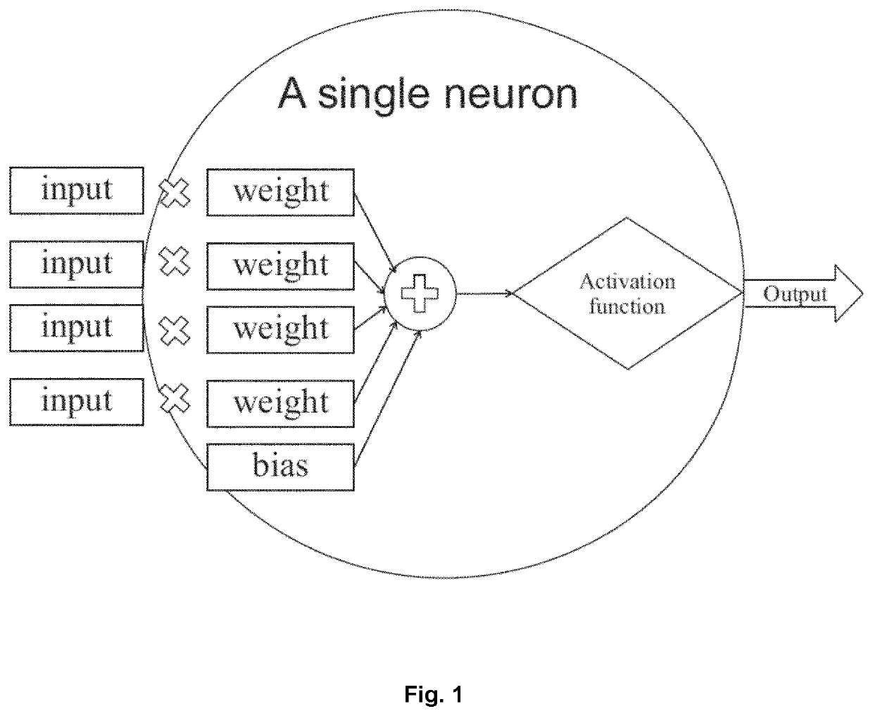
[0134]FIG. 1 illustrates the structure of a neuron as part of a (convolutional) neural network, in which input is assigned certain weights for processing by an activation function which generates the output of the neuron.

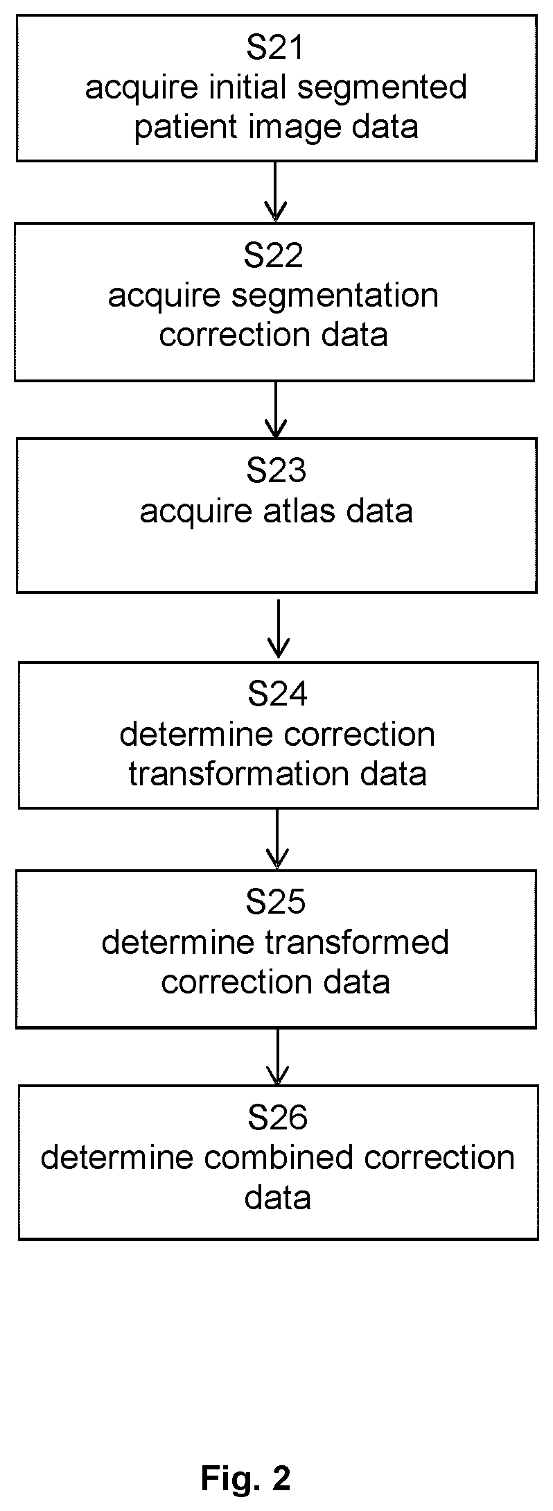
[0135]FIG. 2 describes the basic flow of the method according to the first aspect, which starts in step S21 with acquiring the initial segmented patient image data, continues to step S22 which encompasses acquisition of the segmentation correction data, and then proceeds to acquiring the atlas data in step S23. On that basis, step S24 calculates the correction transformation data, which is followed by determination of the transformed correction data in step S25. Finally, the combined correction data is determined in step S26.

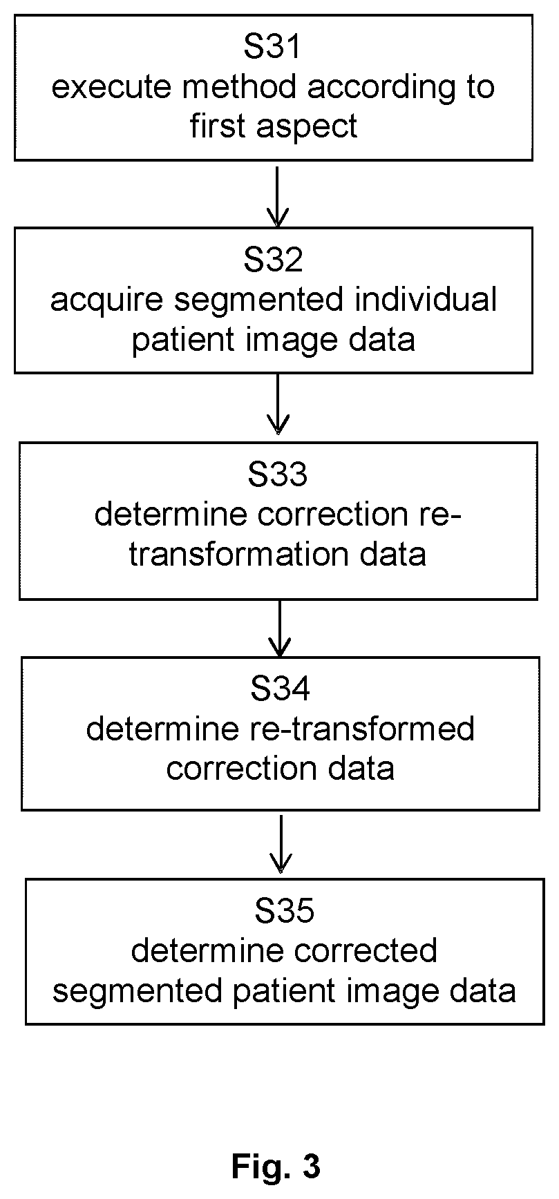
[0136]FIG. 3 illustrates the basic steps of the method according to the second aspect, in which step S31 encompasses execution of the method according to the first aspect and step S32 relates to acquisition of the segmented individual patient im...


PUM


Abstract

Disclosed is a computer-implemented methods of determining distributions of corrections for correcting the segmentation of medical image data, determining corrections for correcting the segmentation of medical image data, training a learning algorithm for determining a segmentation of a digital medical image, and determining a relation between an image representation of the anatomical body part in an individual medical image and a label to be associated with the image representation of the anatomical body part in the individual medical image using the trained machine learning algorithm. The methods encompass reading a plurality of corrections to image segmentations, wherein the corrections themselves may have been manually generated, transforming these corrections into a reference system which is not patient-specific such as an atlas reference system, conducting a statistical analysis of the correction, and applying the re-transformed result of the statistical analysis to patient images. The result of the statistical analysis may also be used to appropriately train a machine learning algorithm for automatic segmentation of patient images. The application of such a trained machine learning algorithm is also part of this disclosure.

Description

FIELD OF THE INVENTION[0001]The present invention relates to a computer-implemented method of determining distributions of corrections for correcting the segmentation of medical image data, methods for correcting the segmentation using the determined distributions of corrections, a corresponding computer program, a computer-readable storage medium storing such a program and a computer executing the program, as well as a medical system comprising an electronic data storage device and the aforementioned computer.TECHNICAL BACKGROUND[0002]It is known to manually generate a correction of image segmentation and to positionally transform the correction into a coordinate space which is not patient-specific, e.g. for reasons of data security. However, this correction by itself provides no reliable measure for quantifying the reliability of the applied segmentation algorithm and does not provide for any consideration of regions of low reliability of the segmentation algorithm during the corr...

Claims


Application Information

Patent Timeline
IPC IPC(8): G06T7/143G06T7/00G06T7/174
CPCG06T7/143G06T7/0012G06T7/174G06T2207/20084G06T2207/20128G06T2207/20092G06T2207/20081G06T2207/30004G06T7/11G06N3/08G06N20/20G06T2207/10116G06T2207/10132G06T2207/10088G06N3/045
Inventor VILSMEIER, STEFANBLUMHOFER, ANDREASSCHMALER, JENS
Owner BRAINLAB
Why Eureka
Try Eureka