
Methods for Improving Intraoral Dental Scans

a dental scan and intraoral technology, applied in the field of intraoral dental scans, can solve the problems of not using artificial intelligence to fill in these missing spots accurately, well the final scan works and how accurate it is, and the accuracy of the produced scan far exceeds the accuracy of physical dental impressions. the effect of improving the intraoral scan

Pending Publication Date: 2022-06-02
LEMCHEN MARC
View PDF0 Cites 0 Cited by
  • Summary
  • Abstract
  • Description
  • Claims
  • Application Information

AI Technical Summary

Benefits of technology

The patent describes a method and apparatus for improving intraoral scans by using data from a reference image to improve the quality of a secondary image captured by a patient. The method involves performing an intraoral scan to produce a reference image, performing a self-intraoral scan to capture a secondary image, and then determining if the secondary image has any missing image data. If there is, the secondary image is improved using data from the reference image. The improved image can then be transmitted to an appliance manufacturer or a processor / server for review and further treatment. The patent also describes a method for providing a visual indicator to the operator to review the secondary image. The technical effects of this patent include improving the accuracy and quality of intraoral scans and providing a more complete record of treatment progress.

Problems solved by technology

While these are amazingly accurate devices, they require trained individuals to operate them when scanning a patient's teeth.
Additionally, because these type of 3D scans require relatively large pieces of equipment and trained operators, the patient must physically travel to an office each time a scan is required which is not only inconvenient but which can further expose the patient and the operator to COVID-19 and other transmittable illnesses.
However, the scans that are produced far exceed the accuracy of physical dental impressions, including those taken by patients at home, often for do-it-yourself aligners or retainers through services such as Smile Direct Club®.
The issue with many of these at home scans however is how well the scan works and how accurate the final scan is.
As of yet, artificial intelligence has not yet been used to fill in these missing spots accurately.

Method used


Image

  • Methods for Improving Intraoral Dental Scans

Examples


Embodiment Construction

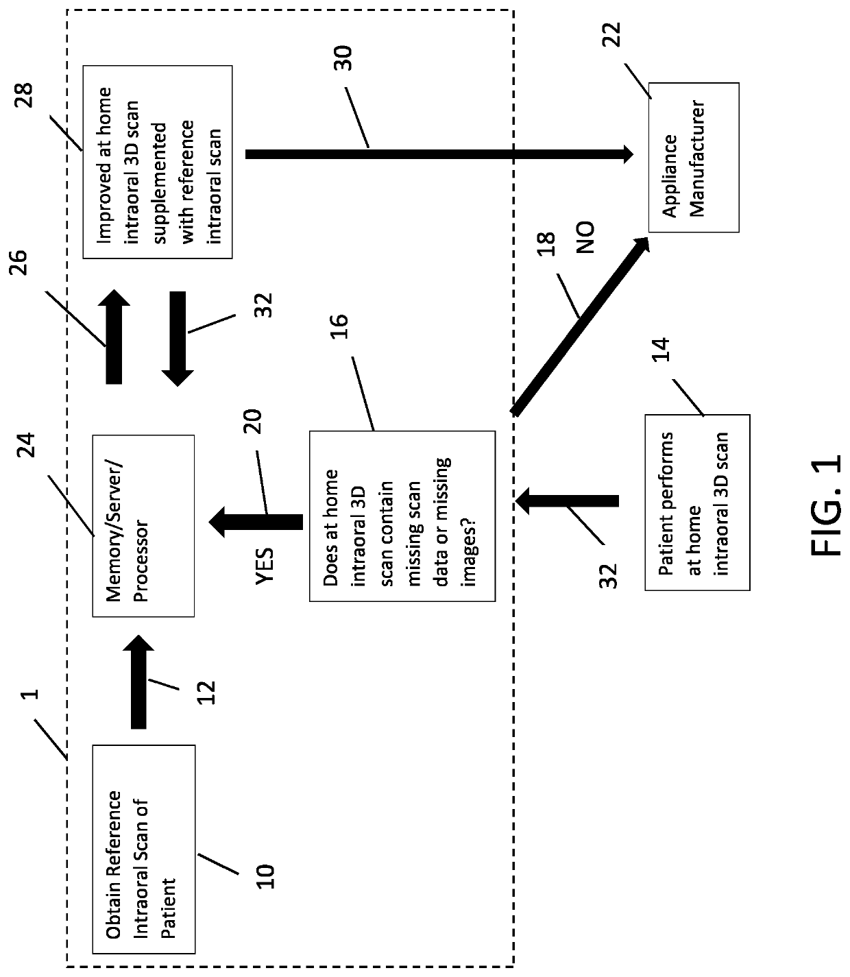
[0025]In a first embodiment of the current invention, a method is provided for improving 3D intraoral scans performed at home which allows the dental professional to directly fill in any missing 3D image data with pre-existing 3D image data that was previously obtained from the patient. Referring to FIG. 1, an operator such as a dentist, orthodontist, or other dental technician performs an in-office highly accurate 3D intraoral scan of a patient using a 3D intraoral scanning system such as the Itero Element® or 3Shape TRIOS® to produce a reference intraoral scan 10. Once completed, the reference 10 scan is sent in step 12 to a memory, server, processor, or other device / location 24 which is accessible by both the operator and the patient via a wireless network such as the internet.

[0026]Next, the patient while at home performs a secondary scan 14 using a camera, smart phone, tablet, or any other smart device which comprises the ability to record an image or a series of images and can...


PUM


Abstract

A method for improving 3D intraoral scans performed at home which allows the dental professional to directly fill in any missing 3D image data with pre-existing 3D image data that was previously obtained from the patient. After the at home intraoral scan is completed, the patient transmits it to the dental professional who then reviews it and determines whether or not the at home intraoral scan comprises any missing 3D image data required for producing a complete and accurate 3D model. If the self-intraoral scan is complete, the dental professional may transmit it directly to an appliance manufacturer. However, if the at home intraoral scan is incomplete and comprises missing 3D image information, the dental professional may instead complete the provided scan by comparing it with the reference intraoral scan. The improved scan is then sent to the appliance manufacturer who may then produce the required appliance.

Description

RELATED APPLICATIONS[0001]The present application is a non-provisional of U.S. provisional application Ser. No. 63 / 119,818, filed on Dec. 1, 2020, which is incorporated herein by reference in its entirety and to which priority is claimed pursuant to 35 USC 119.BACKGROUNDField of the Technology[0002]The invention relates to methods and apparatus for improving intraoral dental scans, specifically a method which allows a patient to produce high quality and complete intraoral dental scans from home which may be used to fabricate aligners, retainers, or other dental or orthodontic appliances.Description of the Prior Art[0003]Currently, when dental appliances such as retainers or a series of aligners are required for a patient, an accurate representation of the patient's teeth is required. Most often this representation is obtained either through a 3D image scan or by taking a physical impression of the patient's teeth.[0004]With regard to 3D image scans, most scans of teeth are done usin...

Claims


Application Information

Patent Timeline
Patent Type & Authority Applications(United States)
IPC IPC(8): A61C9/00G06T7/00G06Q50/04A61C13/00
CPCA61C9/0053G06T7/0014G06T2207/30036A61C13/0004G06Q50/04Y02P90/30G16H30/40G16H30/20G06F9/5077
Inventor LEMCHEN, MARC
Owner LEMCHEN MARC
Learn More