Anti-her2 binding molecules

a technology of anti-her2 and binding molecules, which is applied in the field of binding proteins, can solve the problems of poor outcomes of her2 over-expression and amplification, undefined cell cycle arrest and apoptosis cell death, and the effect of complement dependent cytotoxicity, antigen-dependent cell-mediated cytotoxicity (adcc) and/or complement-dependent cytotoxicity, and improve internalization capability. , the effect of increasing the number of antigen-dependent cell-mediated

Pending Publication Date: 2022-06-02
OLIVIA NEWTON JOHN CANCER RES INST
View PDF0 Cites 0 Cited by
  • Summary
  • Abstract
  • Description
  • Claims
  • Application Information

AI Technical Summary

Benefits of technology

[0304]It may be desirable to modify the antibody of the disclosure with respect to effector function, e.g., so as to enhance antigen-dependent cell-mediated cyotoxicity (ADCC) and / or complement dependent cytotoxicity (CDC) of the antibody. This may be achieved by introducing one or more amino acid substitutions in an Fc region of the antibody. Alternatively, or additionally, cysteine residue(s) may be introduced in the Fc region, thereby allowing interchain disulfide bond formation in this region. The homodimeric antibody thus generated may have improved internalization capability and / or increased complement-mediated cell killing and antibody-dependent cellular cytotoxicity (ADCC). See Caron et al., J. Exp Med. 176:1 191-1 195 (1992) and Shopes, B. J. Immunol. 148:2918-2922 (1992). Homodimeric antibodies with enhanced anti-tumour activity may also be prepared using heterobifunctional cross-linkers as described in Wolff et al. Cancer Research 53:2560-2565 (1993). Alternatively, an antibody can be engineered which has dual Fc regions and may thereby have enhanced complement lysis and ADCC capabilities. See Stevenson et al. Anti-Cancer Drug Design 3:219-230 (1989).
[0305]To increase the serum half-life of the antibody, one may incorporate a salvage receptor binding epitope into the antibody (especially an antibody fragment) as described in U.S. Pat. No. 5,739,277, for example. As used herein, the term “salvage receptor binding epitope” refers to an epitope of the Fc region of an IgG molecule (e.g., IgG1, IgG2, IgG3, or IgG4) that is responsible for increasing the in vivo serum half-life of the IgG molecule. Alternatively, the antibody half-life may be increased by pegylation.
[0306]One form of such an assay is an antigen binding assay, e.g., as described in Scopes (1994) Protein Purification: principles and practice Springer-Verlag. Such a method generally involves labelling the HER2 binding protein and contacting it with immobilized antigen or a fragment thereof, e.g., a protein comprising residues 293 to 309 of mature normal or wild type human HER2 as shown in FIG. 1. Following washing to remove non-specific bound protein, the amount of label and, as a consequence, bound protein is detected. Of course, the HER2 binding protein can be immobilized and the antigen labelled. Panning-type assays can also be used. The examples herein describe binding assays based on flow cytometry.
[0307]HER2 binding proteins that competitively inhibit a HER2 antibody of the invention for binding to an epitope can be screened and identified using conventional competition binding assays known in the art for example, enzyme linked immunosorbent assay (ELISA).
[0308]Assays for determining an HER2 binding protein that competitively inhibits binding of an antibody of the disclosure (e.g. mAb104) will be apparent to the skilled artisan. For example, the antibody of the disclosure is conjugated to a detectable label, e.g., a fluorescent label or a radioactive label. The labelled antibody and the test HER2 binding protein are then mixed and contacted with HER2 or a peptide comprising an epitope thereof (e.g. corresponding to residues 293 to 309 of domain II of human HER2). The level of labelled antibody is then determined and compared to the level determined when the labelled antibody is contacted with HER2 or a peptide comprising an epitope thereof in the absence of the HER2 binding protein. If the level of labelled antibody is reduced in the presence of the test HER2 binding protein compared to the absence of the HER2 binding protein, the HER2 binding protein competitively inhibits binding of the antibody.
[0309]Optionally, the test HER2 binding protein is conjugated to a different label than the antibody. This permits detection of the level of binding of the test HER2 binding protein to the protein or epitope.

Problems solved by technology

HER2 over-expression and amplification has been shown to be associated with poor outcomes in breast and gastric / gastroesophageal junction (GEJ) cancers (Nagaraja V et al., (2016) Eur J Surg Oncol 42(1):140-8); however, its effect on other tumour types is not well defined.
DM1 metabolites disrupt the microtubule networks leading to cell cycle arrest and apoptotic cell death.
Despite the various approaches, there are still challenges.
Despite the successes of Trastuzumab (Herceptin), Pertuzumab and the T-DM1 conjugate, the toxicity of current HER2 antibodies is dose limiting, and resistance invariably develops as discussed above.

Method used


Image

  • Anti-her2 binding molecules
  • Anti-her2 binding molecules
  • Anti-her2 binding molecules

Examples


example 1

Antibody Generation and Characterisation

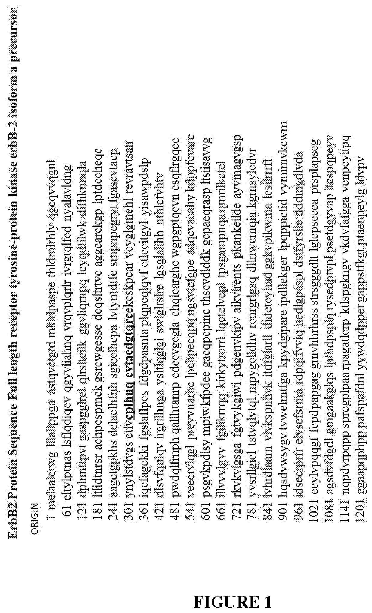
[0441]The immunising antigen and immunisation protocol are described earlier. Through a series of immunisations and screening strategies involving HER2 peptides, recombinant proteins and HER2 expressing cell-based assays, the inventors were finally successful in generating tumour specific monoclonal antibodies to a conformationally flexible region of domain II of HER2.

[0442]The inventors undertook immunization strategies with the linear peptide linked to biotin, GST, MBP, and KLH carrier proteins and Baf / 03 hematopoietic cells (expressing no HER members on cell surface) transfected to express erbB2 with cysteine mutations to expose the peptide loop, but were not successful in generating any clones. Immunisation with the mutant expressing cells plus recombinant mutant ECD ErbB2 did not generate mAbs binding the peptide, but to different locations within the ECD of ErbB2. It was only once the inventors immunized with the cyclized peptide linked ...

example 2

Epitope Analysis and Competition Assays

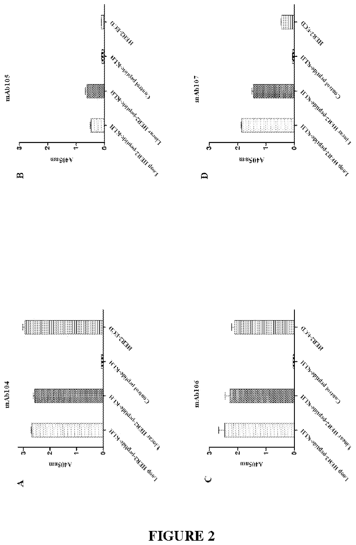
[0459]The mAb104 antibody variable domains binding the antigen epitope located on domain II of HER2 were computationally predicted from homology modelled 3D structures of the antibody Fv domains and the known X-ray structure of human HER2 using the methods previously described (Zhang W, Zeng X, Zhang L, Peng H, Jiao Y, Zeng J, et al. Computational identification of epitopes in the glycoproteins of novel bunyavirus (SFTS virus) recognized by a human monoclonal antibody (MAb 4-5). Journal of Computer-Aided Molecular Design. 2013; 27(6):539-50).

[0460]The predicted HER2 binding of mAb104 was compared with the known crystal structures of Pertuzumab and Trastuzumab binding HER2 (Hu S, Sun Y, Meng Y, Wang X, Yang W, Fu W, et al. Molecular architecture of the ErbB2 extracellular domain homodimer. Oncotarget. 2015; 6(3):1695). Without wishing to be bound by theory, it is thought that the binding of mAb104 to HER2 requires a conformational change that oc...

example 3

Binding of mAb104 to Cell Surface HER2

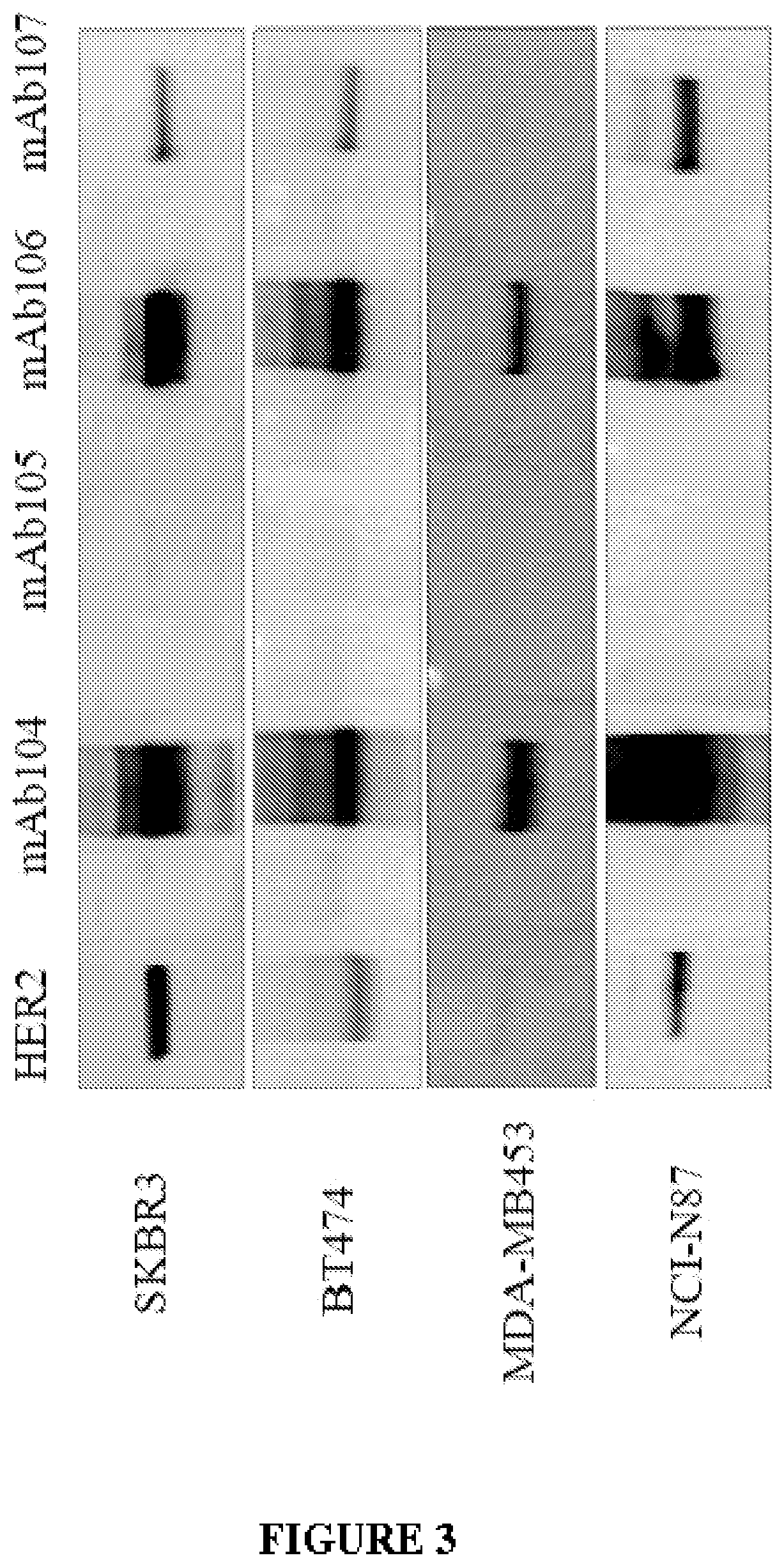
[0472]The inventors examined the pattern and efficiency of mAb104 binding by FACS analysis using a panel of cell lines with differential HER2 expression.

[0473]The results are summarised in Table 8 below. Results are compared to binding with a secondary only antibody.

TABLE 8Binding of mAb104HerceptinPertuzumabmAb104BT-474++++++—SK-BR-3++++++—NCI-N87++++++++OE-19+++++++MDA-MB-231NDND—MCF-7NDND—

[0474]FACS Analysis mAb104 binding to HER2 expressing cells BT474, SK-BR-3, NCI-N87, OE-19, MDA-MB-231, and MCF7 cells were incubated with 10 μg / mL Trastuzumab, Pertuzumab or mAb104 or secondary antibody alone and the extent of binding determined by FACS analysis. Results are representative of two or more experiments

[0475]In cell lines that over-express HER2, mAb104 showed strongest binding to HER2 population in the gastric cell line, NCI-N87, with negligible HER2 binding seen in low HER2-expressing cell lines (MDA-MB-231 and MCF-7).

[0476]Trastuzumab (Hercep...


PUM

PropertyMeasurementUnit
affinity dissociation constantaaaaaaaaaa
concentrationaaaaaaaaaa

Abstract

The present disclosure is directed to binding proteins to the extracellular domain (ECD) of HER2/ErbB2. More particularly, the binding proteins bind to a conformational epitope which is exposed in cells in response to HER2 amplification or activation.

Description

[0001]All documents cited or referenced herein, and all documents cited or referenced in herein cited documents, together with any manufacturer's instructions, descriptions, product specifications, and product sheets for any products mentioned herein or in any document incorporated by reference herein, are hereby incorporated herein by reference in their entirety.[0002]The entire content of the electronic submission of the sequence listing is incorporated by reference in its entirety for all purposes.[0003]This application claims priority to Australian provisional patent application number AU2019900973 filed 22 Mar. 2019. The entire contends of this document are incorporated by reference herein.TECHNICAL FIELD[0004]The present disclosure is directed to binding proteins to the extracellular domain (ECD) of HER2 / ErbB2. More particularly, the binding proteins bind to a conformational epitope which is exposed in cells in response to HER2 amplification or activation.BACKGROUND[0005]The E...

Claims


Application Information

Patent Timeline
Patent Type & Authority Applications(United States)
IPC IPC(8): C07K16/32G01N33/574
CPCC07K16/32A61K2039/505G01N33/57484A61P35/00A61K2121/00G01N33/566A61K2039/507C07K2317/34C07K2317/73C07K2317/92
Inventor SCOTT, ANDREW MARKGAN, HUI KONGLIU, ZHANQIPARAKH, SAGUN
Owner OLIVIA NEWTON JOHN CANCER RES INST
Why Eureka
Try Eureka