Rapid cell culture test device designed such that fluid films with uniform thickness are formed

a cell culture and fluid film technology, applied in biochemistry apparatus and processes, laboratory glassware, instruments, etc., can solve the problems of inability to design the system at the same time, and achieve the effect of accurately and quickly determining the effects of a bioactive agent, the effect of rapid antibacterial susceptibility testing

Pending Publication Date: 2022-11-03
QUANTA MATRIX
View PDF0 Cites 0 Cited by
  • Summary
  • Abstract
  • Description
  • Claims
  • Application Information

AI Technical Summary

Benefits of technology

[0020]The rapid cell culture test device of the present disclosure is designed such that fluid films with a uniform thickness are formed. Due to this design, the height of solid thin films as the fluid films can be maintained constant, allowing for accurate testing compared to conventional antibiotic test devices. The cell culture test device of the present disclosure uses cells immobilized in the solid thin films to observe the behavior of an antibiotic, enabling rapid antibiotic susceptibility testing compared to conventional devices.
[0021]In addition, the rapid cell culture test device of the present disclosure can generally observe changes in the growth of single bacteria within several tens of minutes (usually 30 minutes) and changes in the growth of single colonies of bacteria within 4 to 6 hours. Accordingly, the rapid cell culture test device of the present disclosure can more accurately and rapidly determine the effects of a bioactive agent on a biological agent than conventional devices.
[0022]Furthermore, the rapid cell culture test device of the present disclosure can observe changes of a biological agent responding to a bioactive agent on a single cell colony basis to determine the minimum inhibitory concentration (MIC) or minimum biofilm eradication concentration (MBEC) of the bioactive agent, thus being very useful for biofilm assay as well as antibiotic susceptibility testing.

Problems solved by technology

However, a considerable cost is inevitably involved in designing the system such that the agarose having a viscosity at around its gelling point is quantitatively dispensed at a CV of less than 10%.

Method used


Image

  • Rapid cell culture test device designed such that fluid films with uniform thickness are formed
  • Rapid cell culture test device designed such that fluid films with uniform thickness are formed
  • Rapid cell culture test device designed such that fluid films with uniform thickness are formed

Examples


example 1

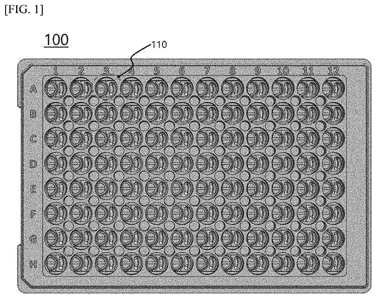
[0087]A thermoplastic resin (polystyrene (PS)) was injection molded into the multi-well-based cell culture test device illustrated in FIG. 1, which had an array of 12×8 aligned well units. After injection molding, the inner walls of the cell culture test device were hydrophilized.

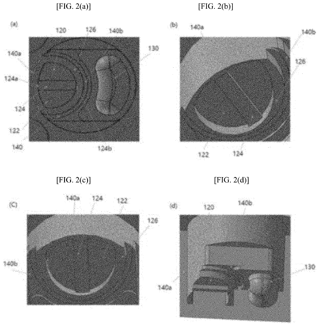
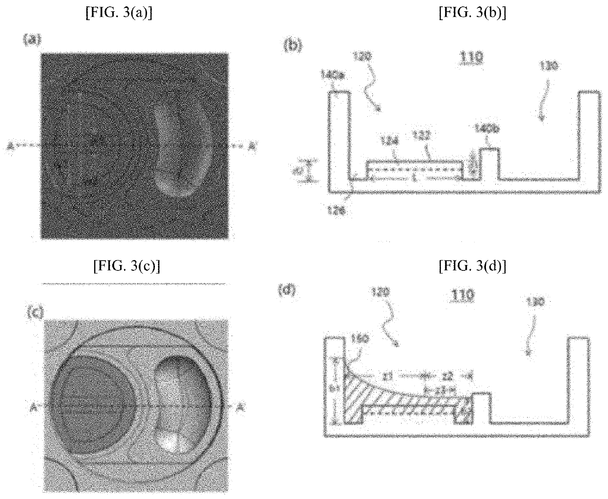
[0088]Agarose containing microbeads was mixed with a bacterial solution (agarose: bacterial solution=50:1) to prepare a first fluid. The first fluid was dispensed into the well units. The agarose solution was evenly spread throughout the first sub-wells 120 by the microchannels 126. The agarose solution was stopped at the same height as the barrier portions B 140b of the barrier structures 140 but rose along the inner walls of the barrier portions A 104a of the barrier structures 140 to a level higher than the surroundings. When the amount of the first fluid was small overall, the width of the capillary channels 124 was made smaller than that of the microchannels 126d, which is preferable in terms of soluti...

example 2

[0093]FIG. 10 shows growth of reference strain E. coli ATCC 25922 without antibiotics, which was observed at regular time intervals using the cell culture test device of the present disclosure. The bacterial colonies increased in size with time. The colonies of the strain were visible to the naked eye from 4 h after initiation of colonization.

[0094]FIG. 11 shows a process for susceptibility testing of reference strain E. coli ATCC 25922 to imipenem as an antibiotic, which was observed at regular time intervals using the cell culture test device of the present disclosure. The antibiotic was used at concentrations of 0.12 μg / ml to 2.0 μg / ml recommended by the Clinical & Laboratory Standards Institute (CLSI). The results were recorded at 2 h intervals until 6 h after initiation of testing. The minimum inhibitory concentration (MIC) of the antibiotic, an important factor in antibiotic susceptibility testing, could be determined, as shown in FIG. 11.

[0095]FIG. 12 shows a process for susc...


PUM

PropertyMeasurementUnit
volumeaaaaaaaaaa
volumeaaaaaaaaaa
volumeaaaaaaaaaa

Abstract

Provided is a multi-well-based rapid cell culture test device designed such that fluid films with a uniform thickness are formed. The rapid cell culture test device has an array structure of a plurality of aligned well units, each of which includes a first sub-well in which a first fluid is accommodated and a barrier structure surrounding the first sub-well to define the area of the first sub-well. A capillary channel recessed from the bottom surface of the first sub-well while traversing the bottom surface is formed to accommodate the first fluid and the barrier structure is divided into a barrier portion A located adjacent to one end of the capillary channel and a barrier portion B located adjacent to the other end of the capillary channel. The barrier portion A is greater in height than the barrier portion B such that when the barrier portion A is wetted with the first fluid introduced into the first sub-well along its inner wall, a change in the level of the first fluid at the other end of the capillary channel depending on the amount of the first fluid dispensed is minimized.

Description

TECHNICAL FIELD[0001]The present disclosure relates generally to a rapid cell culture test device designed such that fluid films with a uniform thickness are formed.BACKGROUND ART[0002]Generally, antibiotic susceptibility testing (response of bacteria to a drug) is performed by placing bacteria in a multi-well plate, introducing a drug in a liquid form, and monitoring time-dependent changes of the bacteria using an optical measurement system. According to the Kirby-Bauer (KB) method for antibiotic susceptibility testing, bacteria are inoculated into a solid medium, placing antibiotic-absorbed papers thereon, and observing bacterial growth. In the case of microdilution testing in liquid media, a number of automated systems such as VITEK2, Microscan, and Phoenix have been developed for antibiotic susceptibility testing. Such a system can be used for antibiotic susceptibility testing by placing an antibiotic in millimeter-sized wells, introducing bacteria and a liquid medium into the w...

Claims


Application Information

Patent Timeline
Patent Type & Authority Applications(United States)
IPC IPC(8): C12Q1/20B01L3/00
CPCC12Q1/20B01L3/50853B01L2200/16B01L2300/0851B01L2300/0829B01L2300/044B01L2300/087C12M23/12B01L3/5085G01N33/5008B01L2200/0647B01L2300/161B01L2300/12
Inventor LEE, GI YOONLIM, TAE GEUNKIM, DONG YOUNG
Owner QUANTA MATRIX
Why Eureka
Try Eureka