Method and device for acquiring a three-dimensional image data set of a moving organ of the body

a three-dimensional image and data set technology, applied in tomography, instruments, applications, etc., can solve the problems of pulsating continuously, not being able to acquire, giving rise to disturbing artifacts, etc., to reduce the number of x-ray positions and the number of projection data sets, and improve image quality.

Inactive Publication Date: 2005-03-08
U S PHILIPS CORP
View PDF6 Cites 93 Cited by
  • Summary
  • Abstract
  • Description
  • Claims
  • Application Information

AI Technical Summary

Benefits of technology

Granted, it is in principle possible to reduce the number of X-ray positions and the number of projection data sets to be acquired and to replace individual projection data sets by interpolated data. However, this always degrades the image quality and such a reduction of the acquired data can be continued to a given lower limit only; below this limit various effects during the data processing, for example the effect of superposition which is due to an insufficient sampling rate, make the formation of a three-dimensional image data set hardly possible or even impossible. Therefore, in accordance with the invention a projection data set of the body organ to be examined is acquired in each X-ray position necessary for the acquisition of a three-dimensional image data set, that is, each time during a low-motion phase of the periodically moving body organ. The periodically moving body organ may at the same time be the body organ to be examined or a body organ which induces a likewise periodic motion of the body organ to be examined (for example, the body organ to be examined may be the brain whose periodic motion is linked to the periodic motion of the heart in such a manner that the brain performs a motion with the same period as the heart). The motion signal acquired at the same time is used to control the X-ray device and the data acquisition according to the invention. The acquisition of the projection data sets need not take place in the sequence of the successively occupied X-ray positions; it is also possible to occupy individual X-ray positions, or all X-ray positions, several times, each time a respective projection data set being acquired and it merely being necessary to ensure that at least one projection data set is acquired during a low-motion phase in each X-ray position.

Problems solved by technology

All of said factors could give rise to disturbing artifacts in the image to be formed.
However, at present it is not possible to acquire during this period of time all the sets of projection data necessary for the acquisition of a three-dimensional image data set, so that it is necessary to inject contrast medium a number of times in succession and to acquire projection data sets successively during a plurality of X-ray cycles.
However, a further problem is then encountered in that the heart pulsates continuously, that is, that it performs an eigenmotion, and that, moreover, the heart is moved to and fro due to the respiratory motion of the patient.
Consequently, the projection data sets acquired contain information on the relevant organ of the body examined in different phases of motion, so that a three-dimensional image data set determined from such projection data sets contains artifacts.
Furthermore, no direct and immediate link is provided between the acquisition of the electrocardiogram and the acquisition of the projection data sets by means of the X-ray device, so that a series of projection data sets which have been acquired during the wrong cardiac motion phase are not evaluated.

Method used


Image

  • Method and device for acquiring a three-dimensional image data set of a moving organ of the body
  • Method and device for acquiring a three-dimensional image data set of a moving organ of the body
  • Method and device for acquiring a three-dimensional image data set of a moving organ of the body

Examples


Embodiment Construction

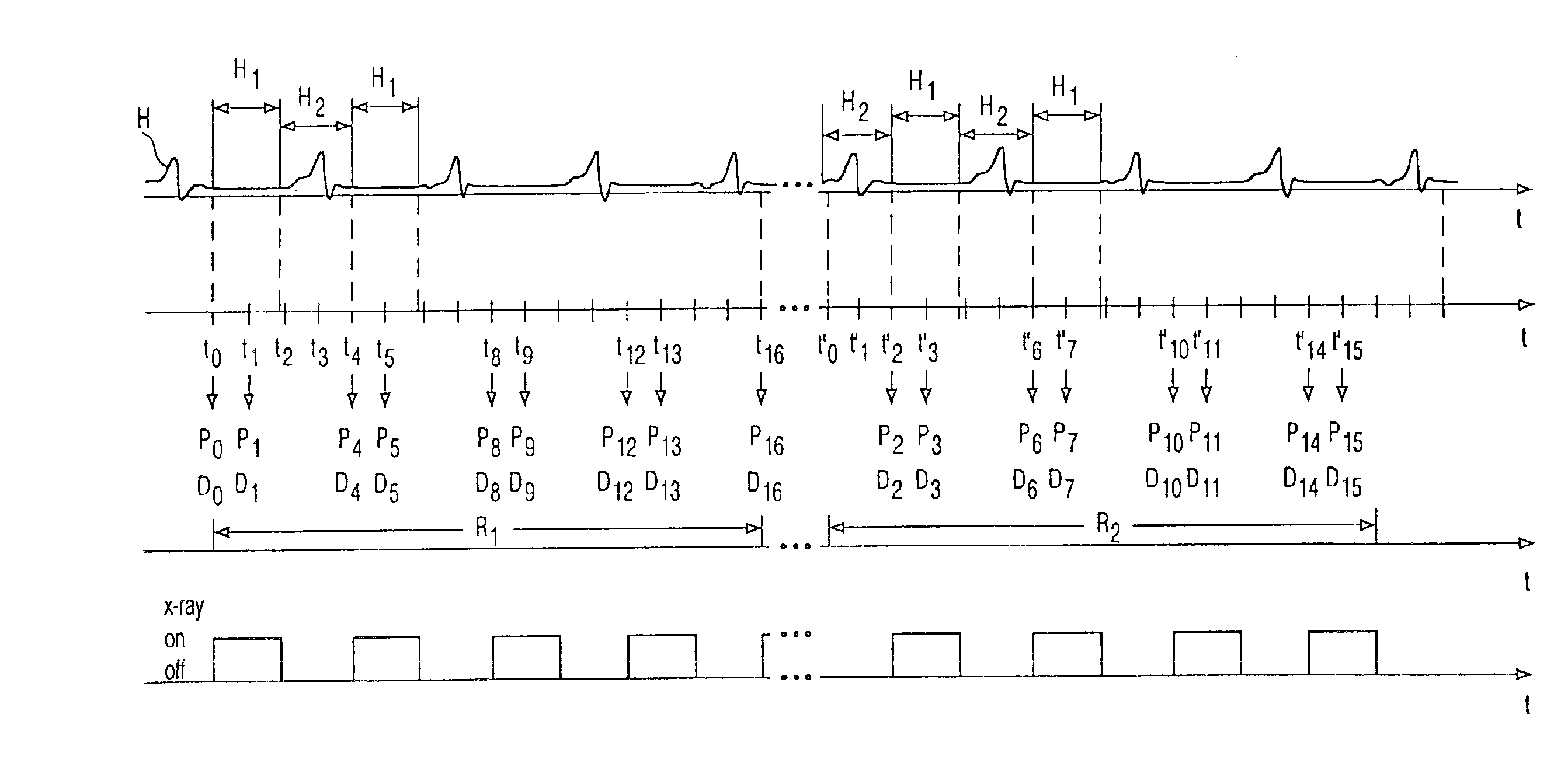
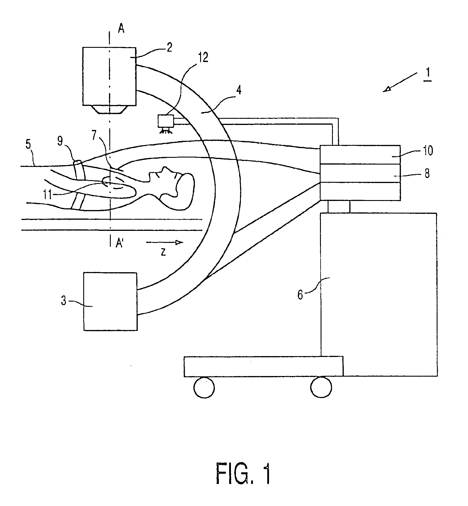
The X-ray device 1 shown in FIG. 1 includes an X-ray tube 2 and a two-dimensional X-ray detector 3, for example an image intensifier, which are mounted on a C-arm 4 in such a manner that they are arranged so as to be rotatable about the z axis and around a patient 5 and can be tilted about an axis extending perpendicularly thereto. The X-ray device 1 and the processing of the data acquired by means of the X-ray detector 3 are controlled by the control and arithmetic unit 6. Electrodes 7 which are connected to an electrocardiography device 8 are arranged on the chest of a patient 5 in order to record an electrocardiogram of the patient. The respiratory motion of the patient 5 is measured by means of an abdominal belt 9 which can be distorted by the respiratory motion and is connected to a respiratory motion measuring device 10 in order to form a respiratory motion signal. The electrocardiogram and the respiratory motion signal are conducted on-line to the control and arithmetic unit ...


PUM

PropertyMeasurementUnit
timeaaaaaaaaaa
phaseaaaaaaaaaa
computed tomographyaaaaaaaaaa

Abstract

The invention relates to a method of and a device for the formation of a three-dimensional image data set of a periodically moving body organ (11) of a patient (5) by means of an X-ray device (1) which includes an X-ray source and an X-ray detector (3), a motion signal (H, B) which is related to the periodic motion of the body organ (11) being measured simultaneously with the acquisition of the projection data sets (D0, D1, . . . , D16). In order to improve such a method or such a device, notably in order to improve the construction and to reduce the time required for data processing while keeping the radiation dose for the patient as small as possible and while ensuring an as high as possible image quality, the invention proposes to acquire the projection data sets (D0, D1, . . . , D16) necessary for the formation of the three-dimensional image data set successively from different X-ray positions (p0, p1, . . . , p16) which are situated in one plane, to control the X-ray device by means of the motion signal (H, B) in such a manner that a projection data set (D0, D1, . . . , D16) is acquired during a low-motion phase of the body organ (11) in each X-ray position (p0, p1, p16) required for the formation of the three-dimensional image data set, and to use the projection data sets (D0, D1, . . . , D16) acquired during the low-motion phase for the formation of the three-dimensional image data set.

Description

BACKGROUND OF THE INVENTIONThe invention relates to a method of acquiring a three-dimensional image data set of a periodically moving organ of the body of a patient by means of an X-ray device in which projection data sets are acquired by the X-ray device simultaneously with a motion signal related to the periodic motion of the organ. The present invention also relates to an X-ray device including an X-ray source and an X-ray detector for acquiring projection data sets from different X-ray positions and enabling the formation of a three-dimensional image data set of a moving organ of the body of a patient from the projection data sets, and a mechanism for measuring a motion signal related to the periodic motion of the organ which is acquired simultaneously with the acquisition of projection data sets.SUMMARY OF THE INVENTIONFor the acquisition of a three-dimensional image data set of an object by means of an X-ray device it is necessary to acquire a plurality of sets of projection d...

Claims


Application Information

Patent Timeline
Patent Type & Authority Patents(United States)
IPC IPC(8): A61B6/00A61B6/02
CPCA61B6/541A61B6/5288A61B6/4441A61B6/032A61B5/02444A61B5/14551A61B5/0809
Inventor RASCHE, VOLKERGRASS, MICHAEL
Owner U S PHILIPS CORP
Why Eureka
Try Eureka