
Microscope, flow cytometer, and method for examination of a specimen

a flow cytometer and microscope technology, applied in the field of microscopes and flow cytometers, can solve the problems of distortion of spectrum, inability to obtain full spectrum, and inability to observe specimens to an even more extreme extent, and achieve the effect of very rapid switching

Inactive Publication Date: 2005-03-15
LEICA MICROSYSTEMS CMS GMBH
View PDF6 Cites 41 Cited by
  • Summary
  • Abstract
  • Description
  • Claims
  • Application Information

AI Technical Summary

Benefits of technology

A further object of the invention is to describe a flow cytometer that makes possible improved, largely uninterrupted and defect-free, high-resolution spectral analysis of the detection light.
The fluorescence resonance energy transfer (FRET) efficiency of specifically labeled specimens can be measured using the method according to the present invention. With the method according to the present invention, the FRET shift (which differs depending on the environment of the structures being examined) can be measured easily and efficiently, permitting conclusions as to the composition and behavior of the specimen under examination.

Problems solved by technology

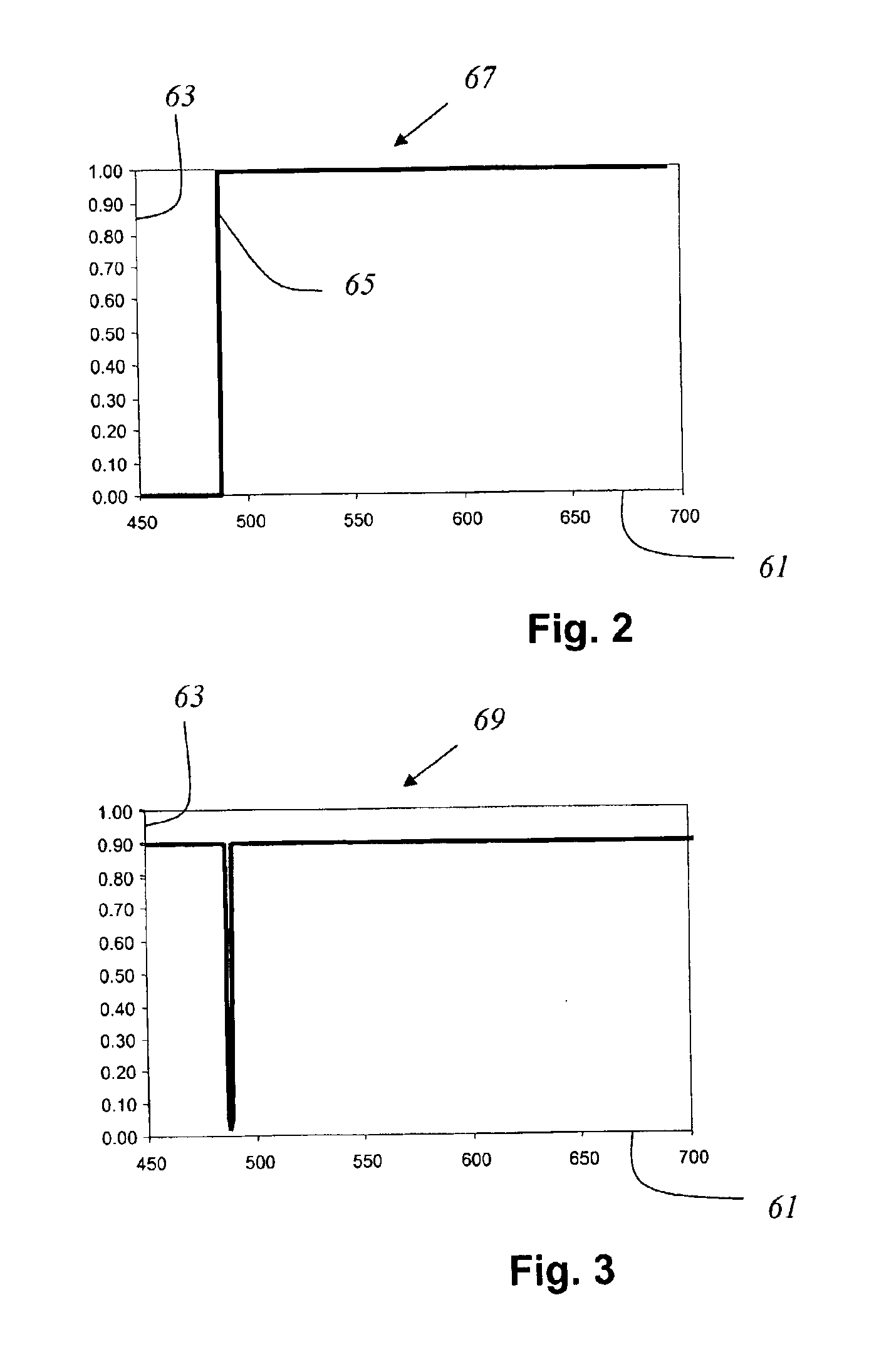
The apparatuses and methods disclosed in the aforesaid documents have the disadvantage that the spectrum acquired in each case, which is generated from the light proceeding from the specimen, is incomplete in a very broad region of several tens of nanometers around the wavelength of the illuminating light.
Because the power level of the fluorescent light is much lower than that of the reflected excitation light, the use of semitransparent neutral splitters to separate the illuminating and detection light also does not solve the aforesaid problem, but merely causes distortion of the spectrum.
The known scanning microscopes and methods prove to be very particularly disadvantageous in applications which involve the analysis of specimens that are labeled with several dyes simultaneously, since in these experiments the illuminating light has two, three, or more wavelengths, so that the aforementioned problems are evident to an even more extreme and troublesome extent.
This scanning microscope has the disadvantage of a very low spectral resolution, since the dispersion properties of usual acoustooptical elements, such as acoustooptical deflectors (AOD) or acoustooptical tunable filters (AOTF) are neither linear nor adequate to achieve sufficient spreading with a reasonable physical size.
The same problems occur analogously with flow cytometers, in which the specimen consists of a fluid shaped into a stream by way of a nozzle.

Method used


Image

  • Microscope, flow cytometer, and method for examination of a specimen
  • Microscope, flow cytometer, and method for examination of a specimen
  • Microscope, flow cytometer, and method for examination of a specimen

Examples


Embodiment Construction

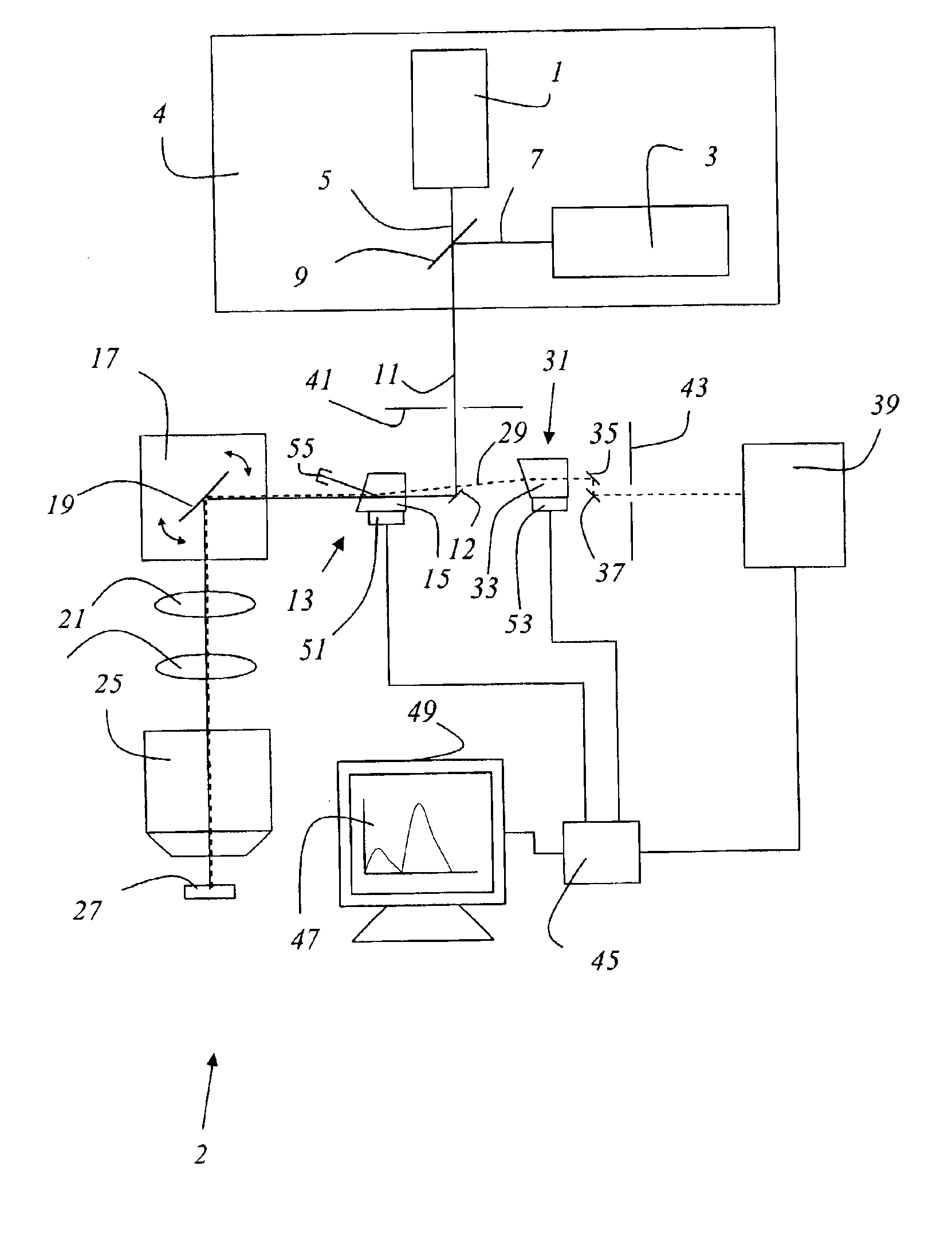
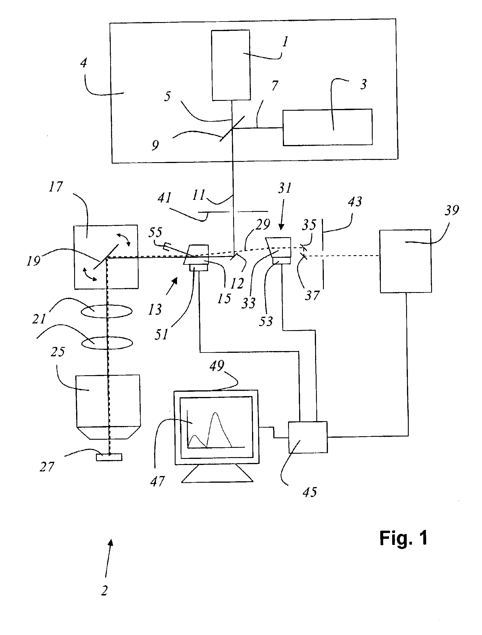
FIG. 1 shows a microscope 2 according to the present invention that is embodied as a confocal scanning microscope, having a light source 4 that contains two lasers 1, 3 whose emitted light beams 5, 7 have different wavelengths, emitted light beams 5, 7 being combined with a dichroic beam combiner 9 into an illuminating light beam 11. The scanning microscope comprises an acoustooptical component 13 that is embodied as an AOTF 15. Illuminating light beam 11 is reflected by a deflecting mirror 12 to acoustooptical component 13. From acoustooptical component 13, illuminating light beam 11 arrives at a beam deflection device 17 that contains a gimbal-mounted scanning mirror 19 and that guides illuminating light beam 11, through scanning optical system 21, tube optical system 23, and objective 25, over or through specimen 27. Detection light beam 29 coming from the specimen travels in the opposite direction through scanning optical system 21, tube optical system 23, and objective 25, and ...


PUM

PropertyMeasurementUnit
excitation wavelengthaaaaaaaaaa
wavelengthsaaaaaaaaaa

Abstract

A microscope with a light source that emits light for illumination of a specimen and with a spectrometer that receives detection light proceeding from the specimen, has an optical arrangement which has an acoustooptical component, and which directs the light of the light source to the specimen and delivers the detection light proceeding from the specimen to a spectrometer in spectrally undivided fashion. A flow cytometer and a method for examination of a specimen are also disclosed.

Description

CROSS REFERENCE TO RELATED APPLICATIONSThis application claims priority of the German patent application 101 62 789.0 which is incorporated by reference herein.FIELD OF THE INVENTIONThe invention concerns a microscope and a flow cytometer.The invention moreover concerns a method for examination of a specimen.BACKGROUND OF THE INVENTIONIn scanning microscopy, a specimen is illuminated with a light beam in order to observe the reflected or fluorescent light emitted by the specimen. The focus of an illuminating light beam is moved in a specimen plane by means of a controllable beam deflection device, generally by tilting two mirrors; the deflection axes are usually perpendicular to one another, so that one mirror deflects in the X direction and the other in the Y direction. Tilting of the mirrors is brought about, for example, by means of galvanometer positioning elements. The power level of the light coming from the specimen is measured as a function of the position of the scanning be...

Claims


Application Information

Patent Timeline
Patent Type & Authority Patents(United States)
IPC IPC(8): G02B21/00
CPCG02B21/0004
Inventor KNEBEL, WERNER
Owner LEICA MICROSYSTEMS CMS GMBH
Learn More