Pre-operative device for localizing marked tissues and process using such a device

a tissue and pre-operative technology, applied in the field of medical techniques for the location of tumors, can solve the problems of difficult ganglions, present devices of nuclear type, not allowing a percentage of detection greater than 80%, and achieve the effect of precise contours

Inactive Publication Date: 2005-07-19
EURORAD 2 6
View PDF7 Cites 28 Cited by
  • Summary
  • Abstract
  • Description
  • Claims
  • Application Information

AI Technical Summary

Benefits of technology

[0019]within the previously determined contours, proceeding with a series of measurements, by the detection of the radioactive radiation emitted by said radioactively marked tissues and / or the detection of the supply or measurement of the magnetic susceptibility of said magnetically marked tissues, so as to determine more precise contours for the zones having peaks of radioactivity and / or magnetic characteristics, and
[0020]producing a second series of reconstituted images of the marked tissues in the zone or zones determined in the preceding step so as to obtain one or more reconstituted images with precise contours of said zone or zones of marked tissues, particularly for the display, temporary storage, editing, filing and / or ultimate processing, for example electronic, of said reconstituted image or images and of the visual expression or expressions of the signal or of the representative signal or signals.

Problems solved by technology

However, the location of the so-called “sentinel” ganglion often gives rise to difficulties, particularly because of its proximity to the principal tumor.
Present devices of the nuclear type, although relatively sensitive, however do not permit a percentage of detection greater than 80%.
However, this second method has several drawbacks connected, on the one hand, to the nature itself of the colorants used (selectivity as to the tumor; retention time; troublesome indelible coloration of the visible tissues because of the necessary concentrations, particularly in cutaneous cancers, in breast cancers; cost .

Method used


Image

  • Pre-operative device for localizing marked tissues and process using such a device
  • Pre-operative device for localizing marked tissues and process using such a device

Examples


first embodiment

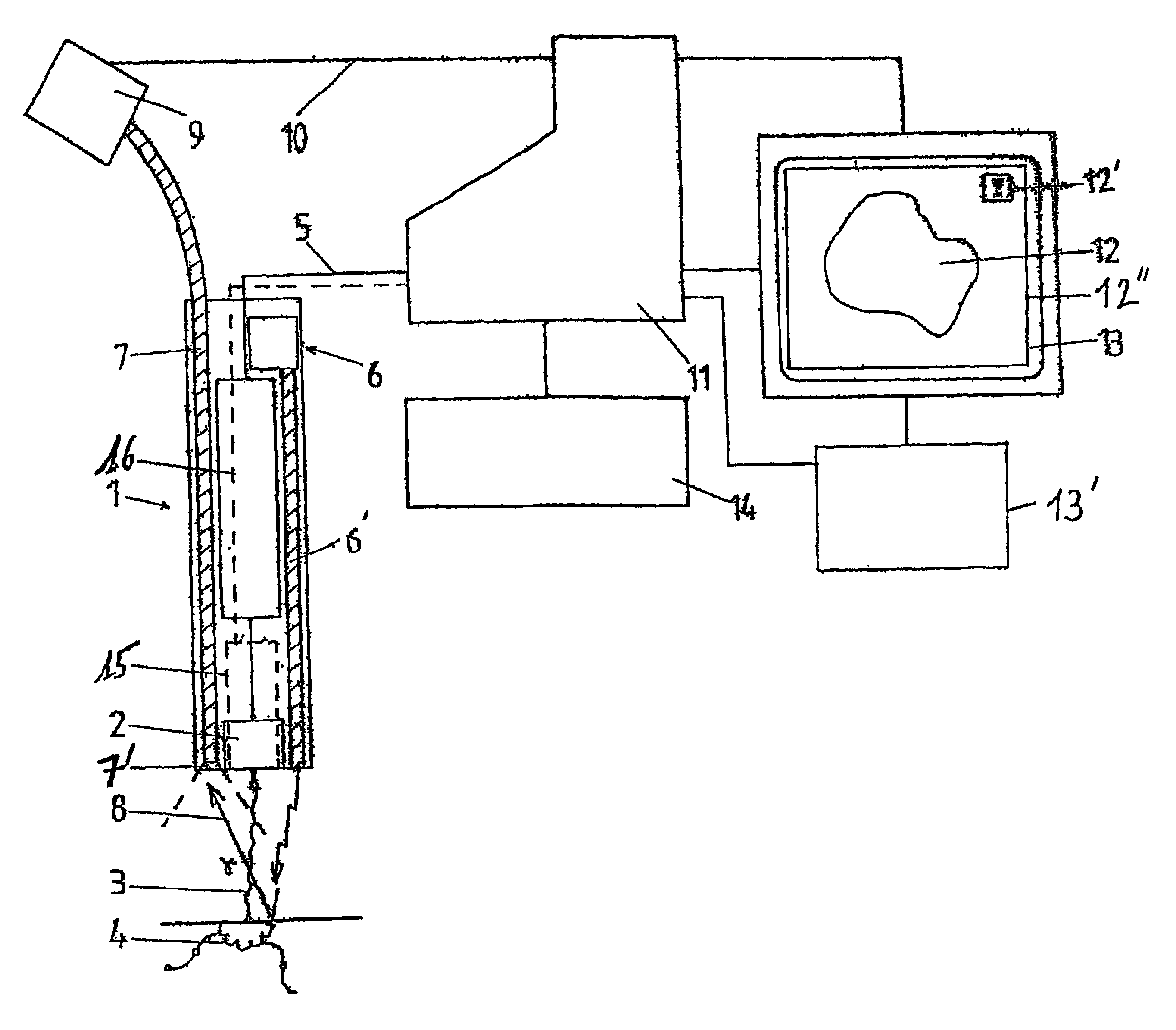
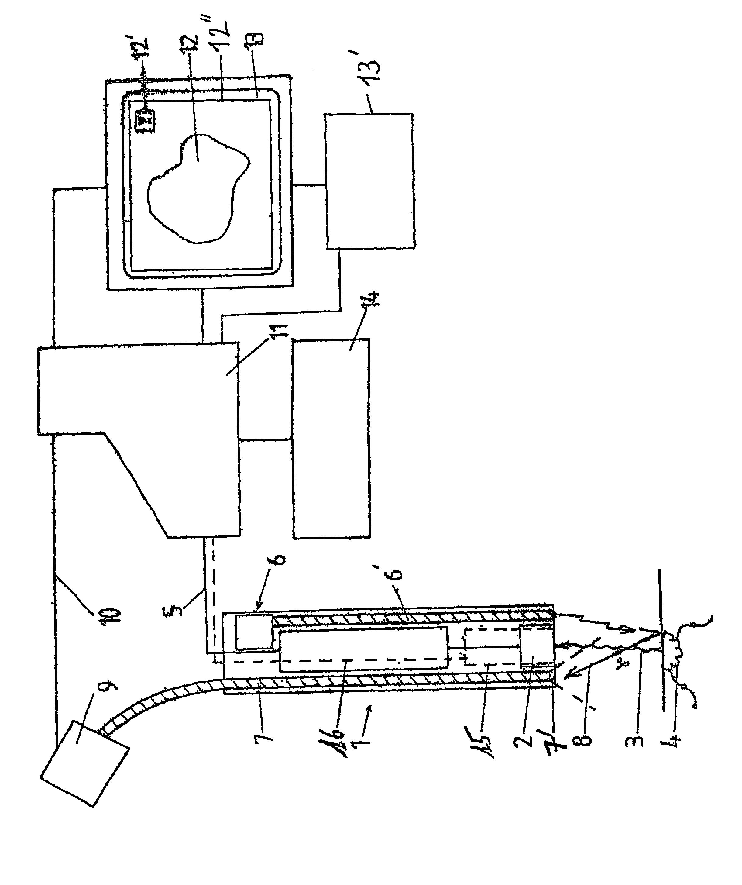
[0030] the probe 1 can comprise a means 2 for the detection of radioactive radiation 3 and at least one optical detection means 6, 7.

second embodiment

[0031] the probe 1 can comprise a means 15 for detecting magnetization or for measuring magnetic susceptibility and at least one optical detection means 6, 7.

[0032]According to a third preferred embodiment of the invention, the probe 1 can comprise a means 2 for detecting radioactive radiation 3, a means 15 for measuring magnetic susceptibility and at least one optical detection means 6, 7.

[0033]The device according to the invention could if desired comprise a set of interchangeable probes 1 corresponding each to one of the above embodiments, each of these probes 1 being if desired used as a function of the difficulty of detection that is anticipated, of the nature of the tissues to be examined or of the type of examination to be carried out. The simultaneous use of means 2, 15 and 6, 7 will permit detection with a very high reliability using three types of detections combined with each other, which is to say associated physically and whose results can be intercorrelated.

[0034]In a ...

second modified embodiment

[0045]According to the invention, the physically perceptible expression 12′ of the representative electric signal or signals 5, 16, simultaneously with the display of reconstituted image 12, takes place in a visual manner, for example by the display or editing of said reconstituted image 12 and if desired the corresponding anatomical image 12″.

[0046]Said visual signal can for example consist in a luminous strip whose filling of the illuminated portion gives a value proportional to the intensity of measured radioactivity or a bi-dimensional image of the distribution of radioactivity in a display or coordinates provided with an origin and axes graduated on the abscissa and ordinate.

[0047]In a particularly practical manner, the physically perceptible expression 12′ of the representative electric signal or signals 5, 16, simultaneously with the display of the reconstituted image 12, takes place on the screen 13 serving to display said reconstituted image 12 and if desired the correspond...


PUM


Abstract

A pre-operative device includes at least one elongated probe (1) integrating at least one element for detecting radioactive radiation (3) emitted by tissues (4) marked by radioactive isotopes or radioactive colloids, generating a first representative electrical signal (5), at least one element (6) for emitting luminous radiation toward the tissues (4) and at least one element (7) for receiving and transmitting reflected luminous radiation (8) reflected by the tissues (4) marked by vital colorants or magnetic markers, toward at least one element (9) for converting the reflected luminous radiation (8) into a second representative electrical signal (10), electrical connection elements for conveying the first and second electrical signals (5, 10) and, at least one electronic means (11) for processing electronic signals.

Description

BACKGROUND OF THE INVENTION[0001]The present invention relates to the field of medical technique for the location of tumors or the like, more particularly devices and probes adapted to be used on or introduced into the human body.[0002]It has for its object a pre-operative location device for marked tissues, particularly by radioactive isotopes of superparamagnetic substances and / or by vital colorants, particularly for the detection and identification of lymphatic ganglia, as well as a process using said device.[0003]The location of marked tissues with the help of isotopes emitting gamma radiation is known for the excision of cancerous tissues as well as for the identification of lymphatic ganglia called “hots”.[0004]The auxiliary ganglia are for example excellent indicators of the prognosis and treatment to be used in the field of melanomas and of breast cancer.[0005]However, the location of the so-called “sentinel” ganglion often gives rise to difficulties, particularly because of...

Claims


Application Information

Patent Timeline
Patent Type & Authority Patents(United States)
IPC IPC(8): A61B5/00G01T1/161
CPCA61B5/0084A61B5/4227A61B6/4258A61B5/4887G01T1/161A61B5/4255A61B5/0086A61B5/4306A61B5/418
Inventor KAZANDJIAN, ANNECHAMBRON, JACQUES
Owner EURORAD 2 6
Why Eureka
Try Eureka