Position tracking and imaging system for use in medical applications

a technology for positioning tracking and imaging system, applied in the field of computer assisted medical surgery, can solve the problems of limiting the movement of the surgical staff, and affecting the accuracy of surgical results

Inactive Publication Date: 2005-08-23
GE MEDICAL SYST GLOBAL TECH CO LLC
View PDF179 Cites 181 Cited by
  • Summary
  • Abstract
  • Description
  • Claims
  • Application Information

AI Technical Summary

Benefits of technology

[0013]In one embodiment, the reference unit is secured from movement with respect to at least a portion of the patient's body such that the reference unit is substantially immobile with respect to a target operation site. The remote unit is attached to the medical instrument. The field generator is associated with one of the reference or...

Problems solved by technology

Such articulated arm tracking systems, however, require that the instrument be attached to a cumbersome mechanical arm.
Also, although free movement of the tip of the arm in three dimensional space may be theoretically possible, the surgeon might experience difficulty positioning the instrument at certain locations and in desired orientations within the body.
This not only limits the movement of the surgi...

Method used


Image

  • Position tracking and imaging system for use in medical applications
  • Position tracking and imaging system for use in medical applications
  • Position tracking and imaging system for use in medical applications

Examples


Embodiment Construction

may be further understood with reference to the accompanying drawings in which:

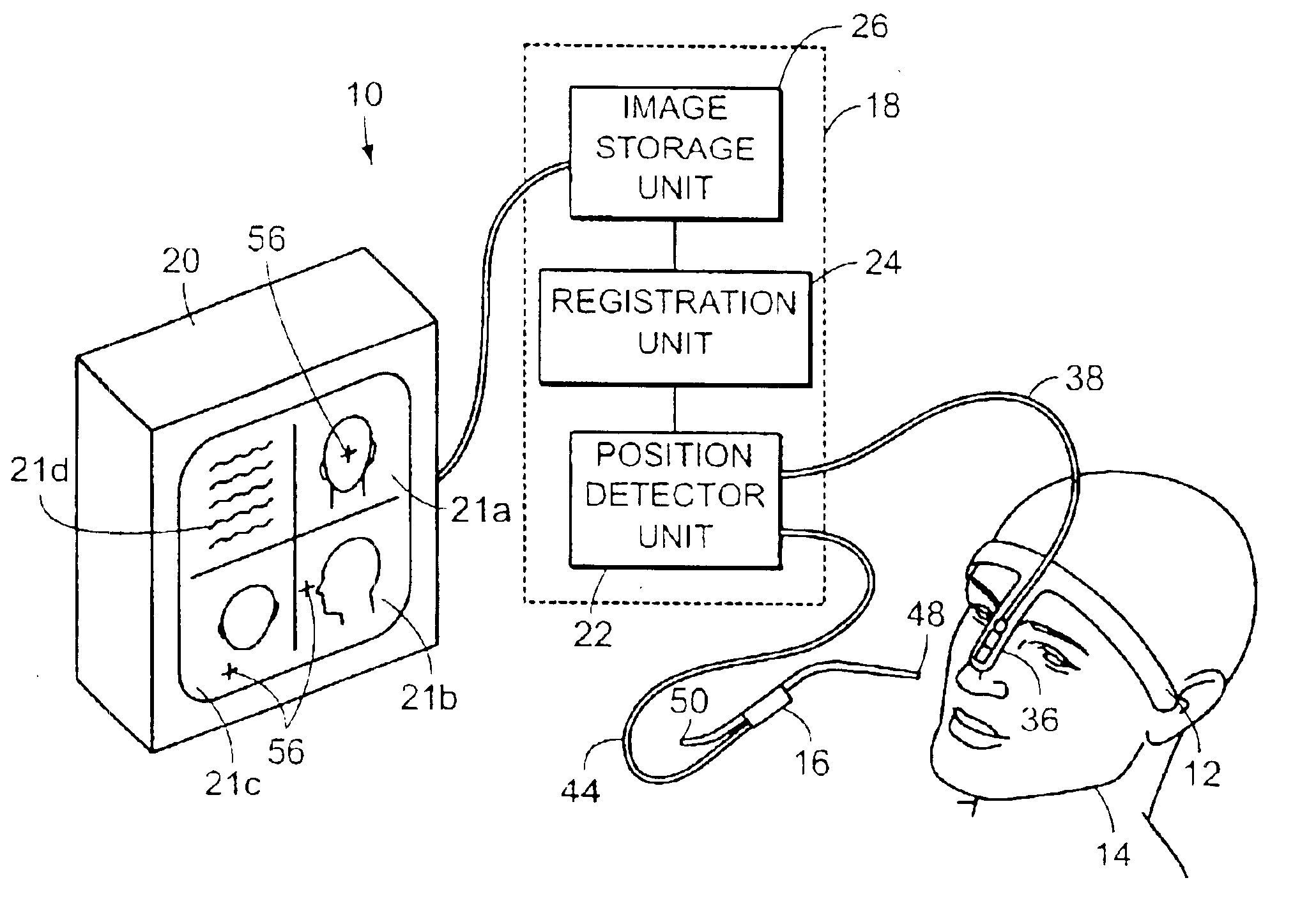
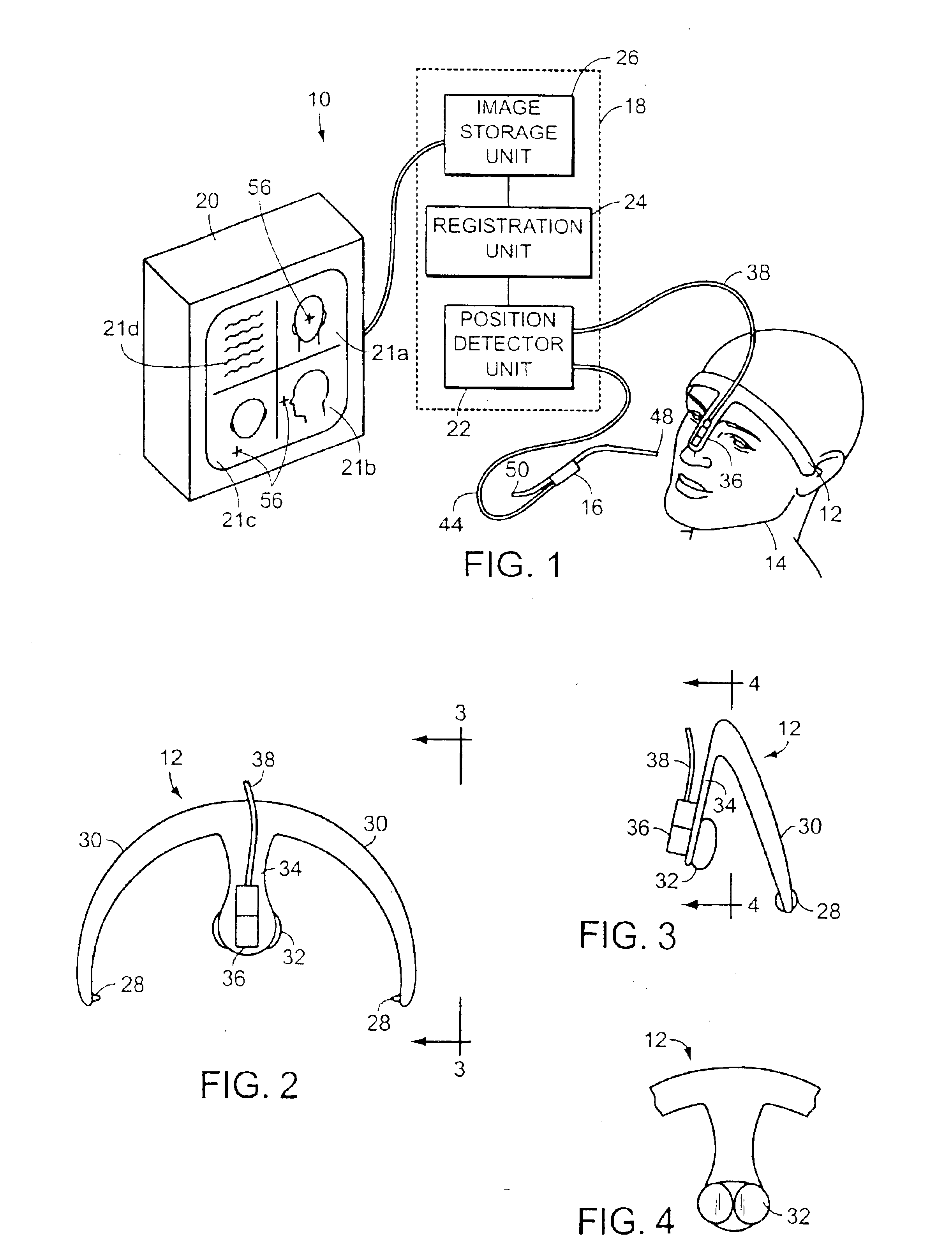
[0018]FIG. 1 is a diagrammatic view of a system of an embodiment of the invention;

[0019]FIG. 2 is a front view of the headset unit shown in FIG. 1;

[0020]FIG. 3 is a side view of the headset unit shown in FIG. 1 taken along line 3—3 of FIG. 2;

[0021]FIG. 4 is a rear view of a portion of the headset shown in FIG. 1 taken along line 4—4 of FIG. 3;

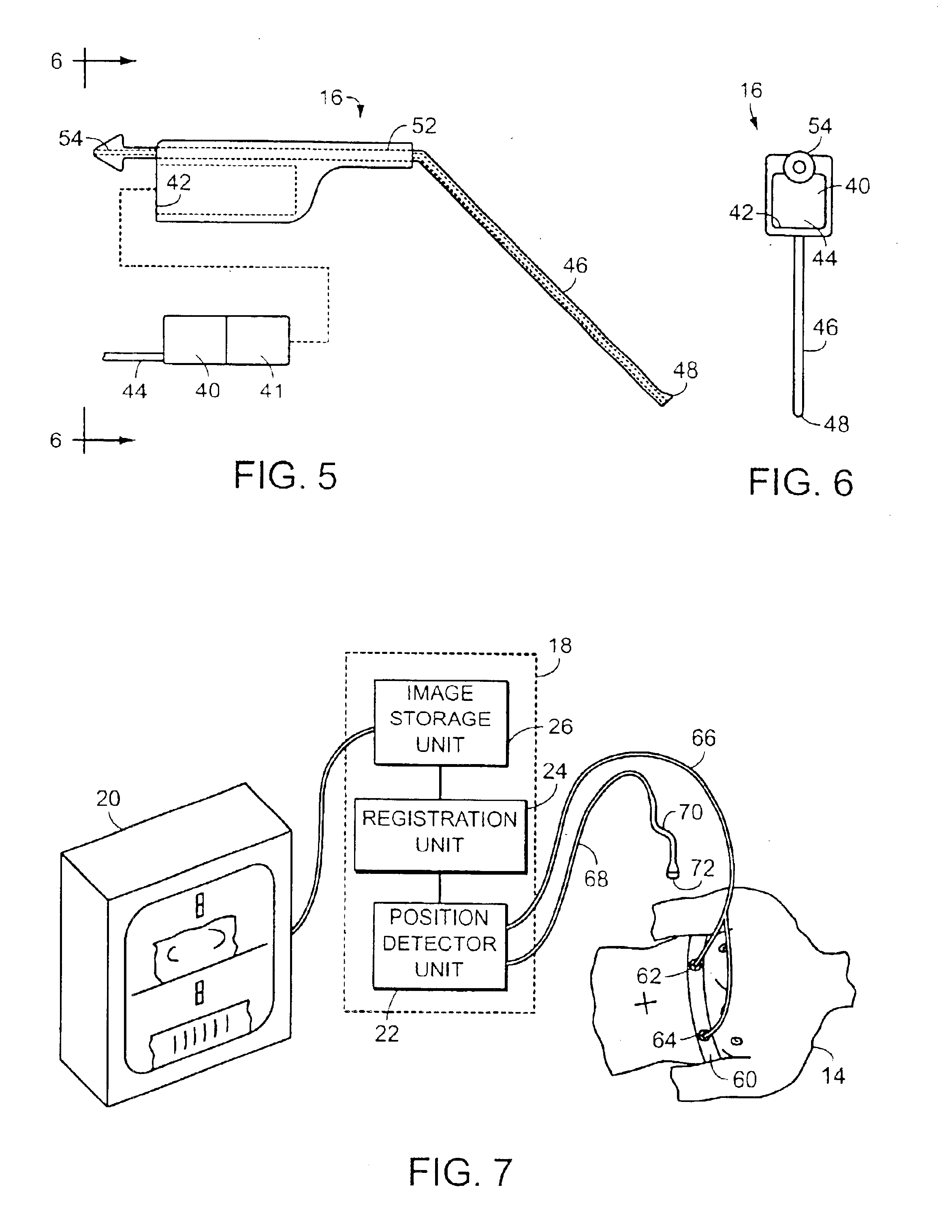
[0022]FIG. 5 is an exploded side view of the surgical instrument and remote sensor shown in FIG. 1;

[0023]FIG. 6 is an end view of the assembled surgical instrument and sensor shown in FIG. 1 taken along line 6—6 of FIG. 5;

[0024]FIG. 7 is a diagrammatic view of an alternate embodiment of the invention;

[0025]FIGS. 8 and 9 are diagrammatic views of image recording and registration operations of the invention; and

[0026]FIGS. 10-13 are diagrammatic views of further embodiments of the invention.

DETAILED DESCRIPTION OF THE ILLUSTRATED EMBODIMENTS

[0027]As shown in FIG. 1, a ...


PUM


Abstract

A method of monitoring a position of a medical instrument with respect to a patient's body, including positioning a patient reference unit on a portion of the patient's body; positioning a remote unit with respect to the medical instrument; associating at least one field sensor with at least one of the patient reference unit and the remote unit; generating an electromagnetic position characteristic field; producing position data responsive to a sensor output signal that is responsive to the presence of the electromagnetic position characteristic field such that the sensor output signal is representative of a position of the remote unit with respect to the patient reference unit; determining image data representing a plurality of images of the patient's body; and displaying an image of the patient's body based on the image data and responsive to position image data that varies for different position data.

Description

[0001]This is a continuation of Ser. No. 09 / 211,365, filed Dec. 14, 1998, now U.S. Pat. No. 6,445,943, issued Sep. 3, 2002, which is a continuation of Ser. No. 08 / 768,305, filed Dec. 17, 1996, now U.S. Pat. No. 5,967,980, issued Oct. 19, 1999, which is a continuation of Ser. No. 08 / 306,818, filed Sep. 15, 1994, now U.S. Pat. No. 5,829,444, issued November 3, 1998.BACKGROUND OF THE INVENTION[0002]The invention relates to computer assisted medical surgery and in particular relates to systems for displaying prerecorded visual images during surgical operations.[0003]Presently available medical imaging techniques such as CAT (Computerized Axial Tomography), MRI (Magnetic Resonance Imaging), and PET (Position Emission Tomography), are known to be helpful not only for diagnostic purposes, but also for providing assistance during surgery. Prerecorded images may be displayed during surgical operations to provide the surgeon with illustrative reference mappings of pertinent portions of a pati...

Claims


Application Information

Patent Timeline
IPC IPC(8): A61B19/00A61B5/06G06T3/00A61B6/03A61B17/00G01B7/004
CPCA61B5/06A61B6/032A61B6/037A61B19/203A61B19/52A61B19/5244A61B19/54G01B7/004G06T3/0068A61B19/20A61B19/5212A61B2017/00022A61B2017/00464A61B2017/00477A61B2017/00482A61B2019/448A61B2019/4894A61B2019/5251A61B2019/5255A61B2019/5268A61B2019/5272A61B2019/5287A61B2019/5437A61B2019/5483A61B5/062A61B5/064A61B34/20A61B90/10A61B90/14A61B90/36A61B90/361A61B90/39A61B90/98A61B2034/2051A61B2034/2055A61B2034/2068A61B2034/2072A61B2090/0818A61B2090/363A61B2090/3937A61B2090/3983
Inventor FERRE, MAURICE R.JAKAB, PETER D.TIEMAN, JAMES S.
Owner GE MEDICAL SYST GLOBAL TECH CO LLC
Why Eureka
Try Eureka