Registration of nuclear medicine images

a nuclear medicine and image technology, applied in the field of diagnostic imaging, can solve the problems of affecting the accuracy of the image,

Inactive Publication Date: 2005-08-30
GE MEDICAL SYST ISRAEL
View PDF22 Cites 44 Cited by
  • Summary
  • Abstract
  • Description
  • Claims
  • Application Information

AI Technical Summary

Benefits of technology

[0023]Thus, in a preferred embodiment of the invention, image acquisition is gated to body rhythms and motions. Preferably, the structural images are also synchronized in the same manner. For example, gated CT images are used as structural images instead of regular CT images when the STET images, are gated. An advantage of combining STET imaging with gating is the ability to correct binned data for patient motion during data acquisition by realignment based on the registration of the images. This corrects for smearing otherwise produced by patient motion. Additionally, data from separate bins is more easily combined.
[0024]Another advantage is the ability to correct organ motion caused by the gated rhythm, by applying a geometric transformation to data acquired based on the phase of the gated rhythm. Yet another advantage is the ability to register transmission images to emission images even when they are not acquired simultaneously. A transmission image of a patient which is gated to body rhythm can be automatically registered to it corresponding gated emission image, since most of the misalignment between the two images is caused by body rhythms which are, in general, repetitive.

Problems solved by technology

One problem with SPECT is that the tissues surrounding the organs being imaged attenuate and scatter the radiation emitted by the radiopharmaceutical, distorting the resulting SPECT images.
However, STET images have two drawbacks.
First, as indicated above, the STET image does not show much structural detail; therefore, it is difficult to pinpoint where the imaged function is occurring in the patient's body.
Many diagnostic imaging methods, in modalities other than nuclear medicine, reveal almost exclusively structure and not function, therefore, it is hard to compare STET images with other types of diagnostic images.
However, if the two STET images are too different, it is not possible to closely compare them because the operator can not match the different parts of the images to each other.

Method used


Image

  • Registration of nuclear medicine images
  • Registration of nuclear medicine images
  • Registration of nuclear medicine images

Examples


Embodiment Construction

[0034]The present invention does not require the use of any specific STET device, and for most devices the invention can be practiced by changes and / or additions in image processing and registration. In addition, it is possible to use the present invention with NON-STET devices, provided that the SPECT and SPTCT images can be registered to each other.

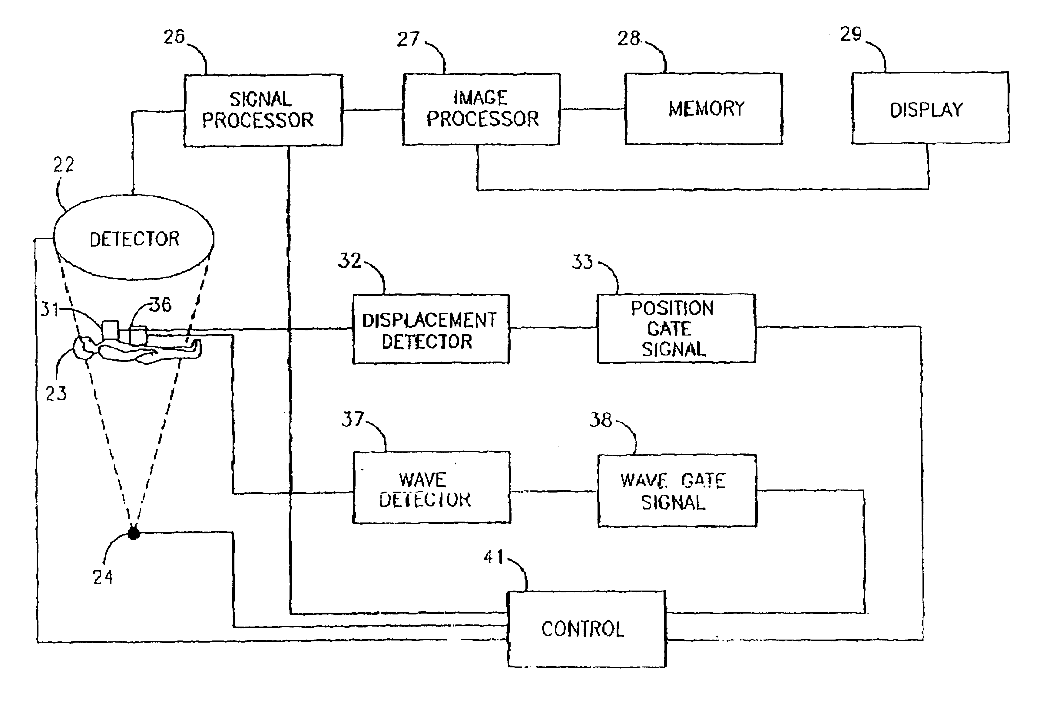
[0035]FIG. 1 in U.S. Pat. No. 5,210,421 shows a typical STET camera assembly which is used for acquiring STET images.

[0036]The process for acquiring these images typically includes:[0037](a) placing a patient on a couch, so that the part to be studied will be in an examination area;[0038](b) injecting a radiopharmaceutical into the patient;[0039](c) acquiring pairs of SPTCT and SPECT images using one or more detectors;[0040](d) rotating the detector(s) around the examination area, in order to acquire a plurality of image pairs;[0041](e) transforming the plurality of image pairs into a multi-slice topographical STET image, a three dimens...


PUM


Abstract

A method of registering a plurality of functional images includes providing a plurality of functional images, providing a plurality of structural images, each one of which having a known positional relationship to at least one of said plurality of functional images, and finding a first mapping transformation between pairs of functional images based on the first mapping transformation and the positional transformation.

Description

RELATED APPLICATIONS[0001]The present application is a divisional of U.S. application Ser. No. 08 / 572,799 filed on Dec. 13, 1995 which is a divisional of U.S. application Ser. No. 08 / 454,871 filed on May 31, 1995 now abandoned and which is related to U.S. application Ser. No. 08 / 571,429 filed on Dec. 13, 1995, now U.S. Pat. No. 5,871,013 issued on Feb. 16, 1999.FIELD OF THE INVENTION[0002]The present invention relates to the art of diagnostic imaging. In particular, the invention relates to nuclear imaging systems incorporating simultaneous transmission and emission tomography or multi-energy window tomography.BACKGROUND OF THE INVENTION[0003]SPECT (Single Photon Emission Computerized Tomography) is used to study the three dimensional distribution of a radionuclide in a patient. Typically one or more radiopharmaceuticals are ingested or are injected into the patient. When radiopharmaceuticals are injected it is usually into the patient's blood stream, to image the cardio-vascular sy...

Claims


Application Information

Patent Timeline
Patent Type & Authority Patents(United States)
IPC IPC(8): G01T1/164G01T1/00G06T3/00G06T7/00G06V10/24
CPCG01T1/1615G01T1/1648G06K9/32G06T3/0068G06T7/0024G06K2209/05G06T2207/30004G06T7/30G06V10/24G06V2201/03
Inventor NATANZON, ALEXBERLAD, GIDEON
Owner GE MEDICAL SYST ISRAEL
Why Eureka
Try Eureka