Breakable gantry apparatus for multidimensional x-ray based imaging

a multi-dimensional computed tomography and apparatus technology, applied in tomography, instruments, applications, etc., can solve the problems of hindering the acceptance and use of mobile three-dimensional imaging in settings outside the radiology department, and the inability to laterally access patients and acquire quality images, so as to facilitate the approach of patients and achieve minimal disruption of medical procedures, high-quality images

Inactive Publication Date: 2005-09-06
MEDTRONIC NAVIGATION INC
View PDF72 Cites 332 Cited by
  • Summary
  • Abstract
  • Description
  • Claims
  • Application Information

AI Technical Summary

Benefits of technology

[0008]An advantage of the present invention relative to conventional CT scanning devices is the ability to manipulate the x-ray gantry around the object to be scanned, and then close the gantry to perform x-ray imaging. For instance, during a medical procedure, a mobile x-ray gantry device of the present invention can easily approach a patient (or be approached by a patient) from a lateral direction, enclose around the patient, and acquire high-quality images (such as three-dimensional x-ray CT images) with minimal disruption of the medical procedure (e.g. to anesthesia, patient monitoring, sterilization, scrub nurses, etc.). In contrast to larger, fixed-bore devices commonly used in hospital radiology departments, the apparatus of the present invention can advantageously be employed in numerous environments, such as operating rooms, procedure rooms, intensive care units, emergency departments and other parts of the hospital, in ambulatory surgery centers, physician offices, and the military battlefield.
relative to conventional CT scanning devices is the ability to manipulate the x-ray gantry around the object to be scanned, and then close the gantry to perform x-ray imaging. For instance, during a medical procedure, a mobile x-ray gantry device of the present invention can easily approach a patient (or be approached by a patient) from a lateral direction, enclose around the patient, and acquire high-quality images (such as three-dimensional x-ray CT images) with minimal disruption of the medical procedure (e.g. to anesthesia, patient monitoring, sterilization, scrub nurses, etc.). In contrast to larger, fixed-bore devices commonly used in hospital radiology departments, the apparatus of the present invention can advantageously be employed in numerous environments, such as operating rooms, procedure rooms, intensive care units, emergency departments and other parts of the hospital, in ambulatory surgery centers, physician offices, and the military battlefield.

Problems solved by technology

The inability to laterally access a patient and acquire quality images has hindered the acceptance and use of mobile three-dimensional imaging in settings outside of the radiology department.

Method used


Image

  • Breakable gantry apparatus for multidimensional x-ray based imaging
  • Breakable gantry apparatus for multidimensional x-ray based imaging
  • Breakable gantry apparatus for multidimensional x-ray based imaging

Examples


Embodiment Construction

[0025]A description of preferred embodiments of the invention follows.

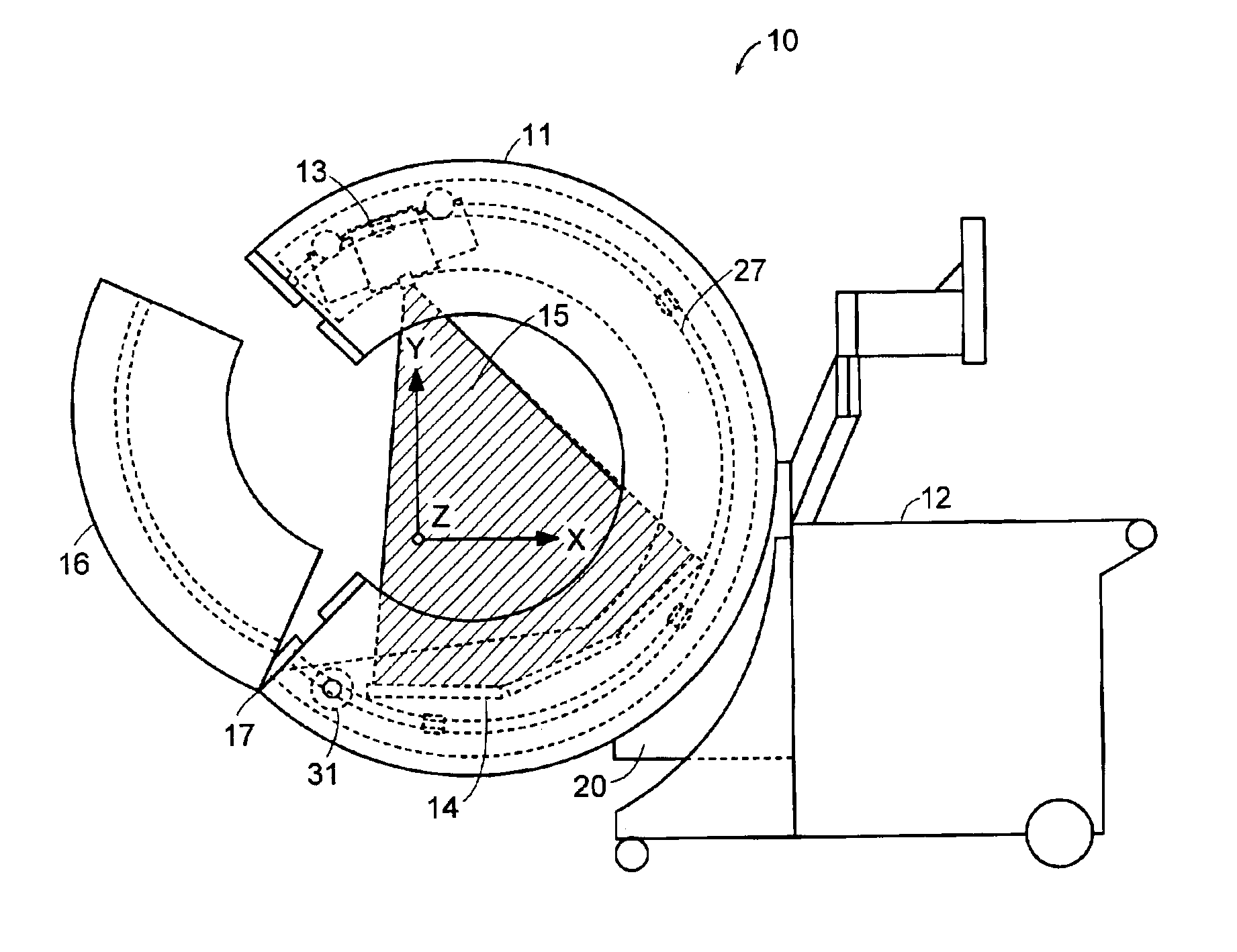
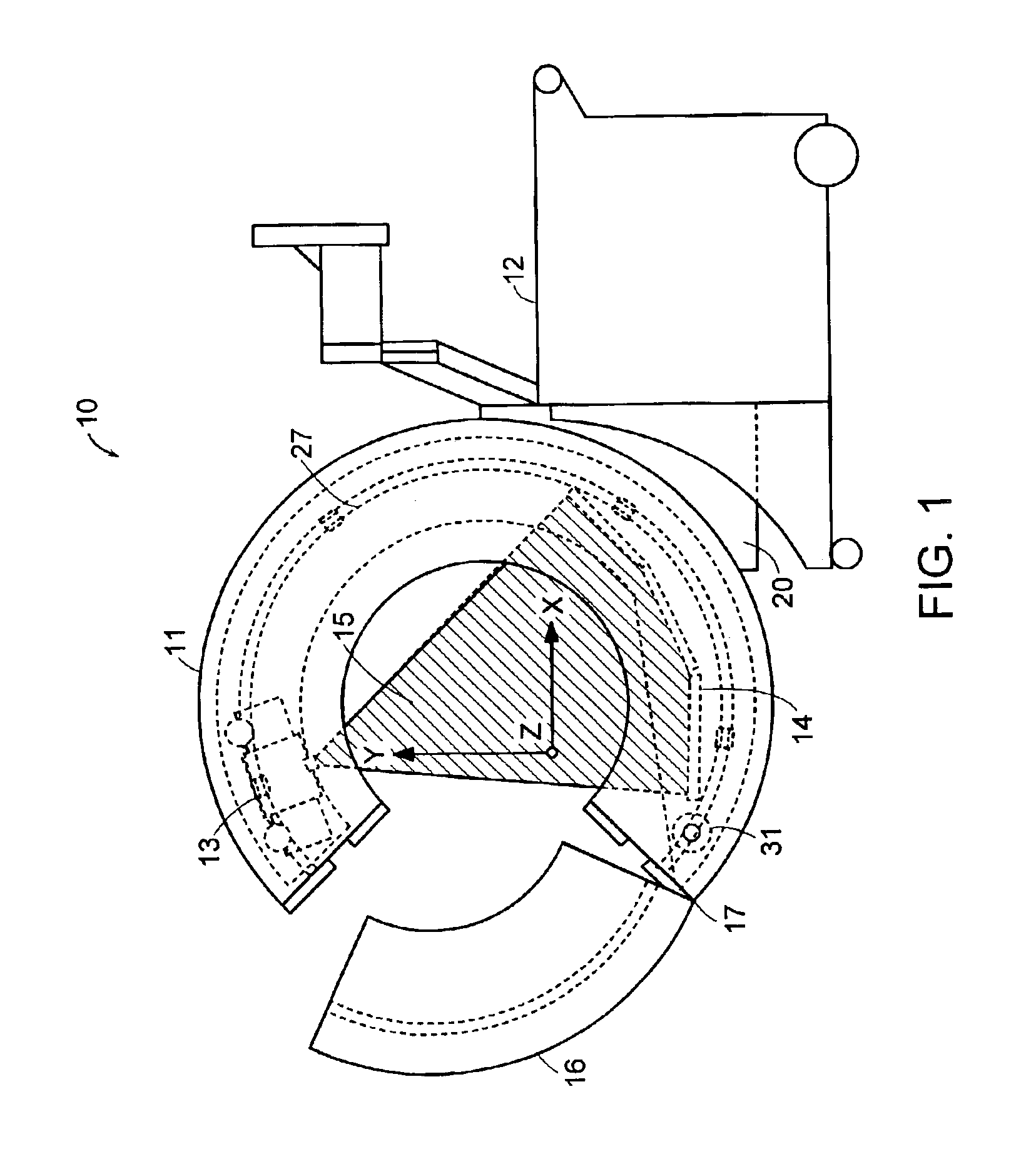
[0026]FIG. 1 is a schematic diagram showing an x-ray scanning system 10, such as a computerized tomographic (CT) x-ray scanner, in accordance with one embodiment of the invention. The x-ray scanning system 10 generally includes a gantry 11 secured to a support structure, which could be a mobile or stationary cart, a patient table, a wall, a floor, or a ceiling. As shown in FIG. 1, the gantry 11 is secured to a mobile cart 12 in a cantilevered fashion via a ring positioning unit 20. In certain embodiments, the ring positioning unit 20 enables the gantry 11 to translate and / or rotate with respect to the support structure, including, for example, translational movement along at least one of the x-, y-, and z-axes, and / or rotation around at least one of the x- and y-axes. X-ray scanning devices with a cantilevered, multiple-degree-of-freedom movable gantry are described in commonly owned U.S. Provisional Applications ...


PUM


Abstract

An x-ray scanning imaging apparatus with a generally O-shaped gantry ring, which has a segment that fully or partially detaches (or “breaks”) from the ring to provide an opening through which the object to be imaged may enter interior of the ring in a radial direction. The segment can then be re-attached to enclose the object within the gantry. Once closed, the circular gantry housing remains orbitally fixed and carries an x-ray image-scanning device that can be rotated inside the gantry 360 degrees around the patient either continuously or in a step-wise fashion. The x-ray device is particularly useful for two-dimensional and / or three-dimensional computed tomography (CT) imaging applications.

Description

RELATED APPLICATION[0001]This application claims the benefit of U.S. Provisional Application No. 60 / 357,398, filed Feb. 15, 2002, the entire teachings of which are incorporated herein by reference.BACKGROUND OF THE INVENTION[0002]Healthcare practices have shown the tremendous value of three-dimensional computed tomographic imaging, mainly as a diagnostic tool in the Radiology Department. Other areas of care, including the operating room, intensive care departments and emergency departments, rely on two-dimensional imaging (fluoroscopy, ultrasound, 2-D mobile X-ray) as the primary means of diagnosis and therapeutic guidance. A truly mobile and practical solution for ‘non-radiology department’ and patient-centric 3-D imaging does not exist. This is mainly due to the fact that current tomographic scanners contain a fixed bore into which the patient must enter from the head or foot. The inability to laterally access a patient and acquire quality images has hindered the acceptance and us...

Claims


Application Information

Patent Timeline
Patent Type & Authority Patents(United States)
IPC IPC(8): A61B6/03A61B6/00
CPCA61B6/032A61B6/4405A61B6/4411A61B6/035A61B6/4441
Inventor GREGERSON, EUGENE A.GRANT, RICHARD K.JOHNSON, NORBERT
Owner MEDTRONIC NAVIGATION INC
Why Eureka
Try Eureka