Vessel detection by mean shift based ray propagation

a mean shift and ray propagation technology, applied in the field of computer vision and imaging systems, can solve the problems of increasing the number of fundamental difficulties, unable to perform topological changes, and the use of snake or balloon methods requires extensive user interaction, so as to enhance ct and mr images, the effect of robustness and efficiency

Inactive Publication Date: 2005-09-20
SIEMENS MEDICAL SOLUTIONS USA INC
View PDF3 Cites 45 Cited by
  • Summary
  • Abstract
  • Description
  • Claims
  • Application Information

AI Technical Summary

Benefits of technology

[0013]A robust and efficient method and system for the segmentation of vessel cross-sections in contrast enhanced CT and MR images is provided. The present invention utilizes boundary propagation by mean shift analysis combined with a smoothness constraint. Consequently, the robustness of the mean shift to noise is further enhanced by the use of a priori information on boundary smoothness. Additionally, the incorporation of the smoothness constraint into the method and system of the present invention allows boundary finding in the presence of eccentricities like calcifications (CT) or small branching vessels. The present invention allows real-time segmentation of medical structures found in multi-modality images (CT, MR, etc.).

Problems solved by technology

However, a number of fundamental difficulties remain; in particular, snakes heavily rely on a proper initialization close to the boundary of an object of interest and multiple initializations are required when multiple objects are to be segmented, i.e., one per object of interest.
However, the balloon model, like snakes, cannot perform topological changes, i.e., balloons can not merge with other balloons and balloon can not split.
Since snakes and balloons cannot easily capture topological changes, the snake or balloon methods require extensive user interaction for images with multiple objects.
A key disadvantage of the level set method is its high computational complexity, due to the additional embedding dimensional, even when the computation is restricted to a narrow band around the curve.
However, these methods are still not fast enough for real-time image segmentation.

Method used


Image

  • Vessel detection by mean shift based ray propagation
  • Vessel detection by mean shift based ray propagation
  • Vessel detection by mean shift based ray propagation

Examples


Embodiment Construction

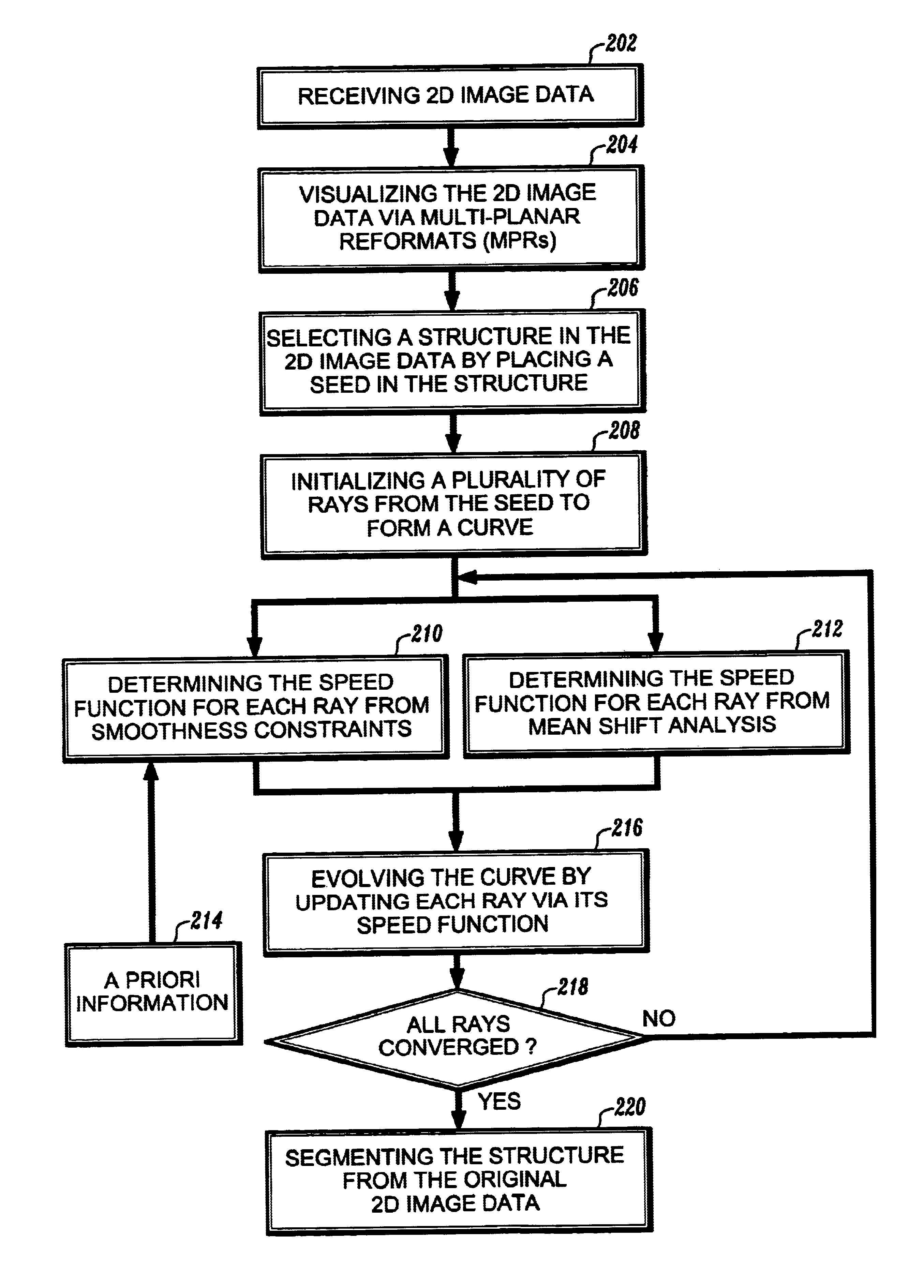
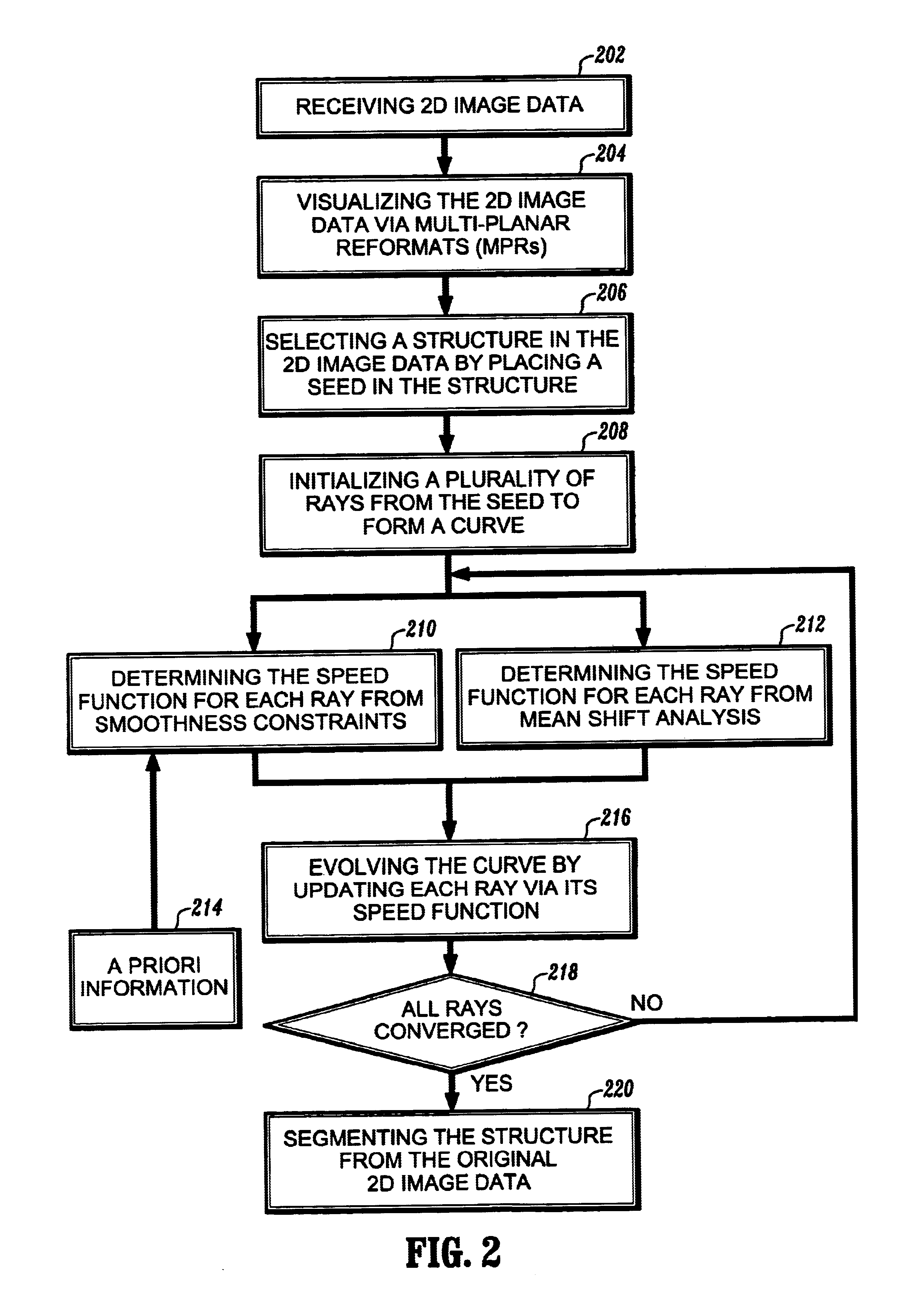
[0033]Preferred embodiments of the present invention will be described hereinbelow with reference to the accompanying drawings. In the following description, well-known functions or constructions are not described in detail to avoid obscuring the invention in unnecessary detail.

[0034]A basic problem in the segmentation of vessels from the background of an image is accurate edge localization, i.e., finding a boundary of the vessel, in the presence of noise. Most CT and MR images have significant noise levels. The present invention employs one dimensional mean shift analysis along a set of rays projecting radially from a user-placed seed point in the image. Noise along these rays is eliminated while edges are preserved. The envelope of these rays represents an evolving contour or curve which converges to the vessel lumen boundary. The method is parsimonious, examining only pixels along and adjoining these rays and the seed point.

[0035]It is important that the boundary determined by th...


PUM


Abstract

A method for segmentation of 2D structures in CT and MR images is provided. The method is based on 2D ray propagation by mean-shift analysis with a smoothness constraint. Ray propagation is used to guide an evolving curve due to its computational efficiency and shape priors are incorporated for robust convergence. The method includes the steps of receiving 2D image data; visualizing the 2D image data on a display device; selecting a structure in the 2D image data by placing a seed in the structure; initializing a plurality of rays from the seed to form a curve; determining a speed function of each of the rays; evolving the curve by propagating the rays based on the speed function of each of the rays; converging the rays on a boundary of the structure; and segmenting the structure when all of the rays have converged on the structure's boundary.

Description

PRIORITY[0001]This application claims priority to an application entitled “VESSEL DETECTION BY MEAN SHIFT BASED RAY PROPAGATION” filed in the United States Patent and Trademark Office on Oct. 23, 2001 and assigned Ser. No. 60 / 347,915, the contents of which are hereby incorporated by reference.BACKGROUND OF THE INVENTION[0002]1. Field of the Invention[0003]The present invention relates generally to computer vision and imaging systems, and more particularly, to a system and method for segmentation of 2D medical structures by mean shift based ray propagation. More specifically, the present invention relates to segmentation of vessel cross-sections in contrast enhanced CT (computerized tomography) and MR (magnetic resonance) images.[0004]2. Description of the Related Art[0005]Images produced by contrast-enhanced magnetic resonance angiography (CE-MRA) and computed tomography angiography (CTA) are becoming an increasingly important tool in the field of medicine. In the CE-MRA imaging pro...

Claims


Application Information

Patent Timeline
Patent Type & Authority Patents(United States)
IPC IPC(8): G06T5/00
CPCG06T7/0012G06T7/0083G06T7/0089G06T2207/10081G06T2207/10088G06T2207/20101G06T2207/20168G06T2207/30101G06T7/12G06T7/149
Inventor TEK, HUSEYINCOMANICIU, DORINWILLIAMS, JAMES
Owner SIEMENS MEDICAL SOLUTIONS USA INC
Why Eureka
Try Eureka