Ultrasonic imaging device

Inactive Publication Date: 2005-10-25
HITACHI MEDICAL CORP
View PDF2 Cites 7 Cited by
  • Summary
  • Abstract
  • Description
  • Claims
  • Application Information

AI Technical Summary

Benefits of technology

[0010]The first object of the present invention is to provide an ultrasonic imaging apparatus tha

Problems solved by technology

But, the structure of the medium to be examined is not uniform.
When the actual sonic velocity is exceedingly different from posited velocity due to individual differences, a clear image cannot be obtained since ideal focusing is not performed.
Heisei 6-269447, however, the medium composition has to be hypothesized and also the calculation method is complicated.
Moreover, the long time it takes to do this calculati

Method used


Image

  • Ultrasonic imaging device
  • Ultrasonic imaging device
  • Ultrasonic imaging device

Examples


Embodiment Construction

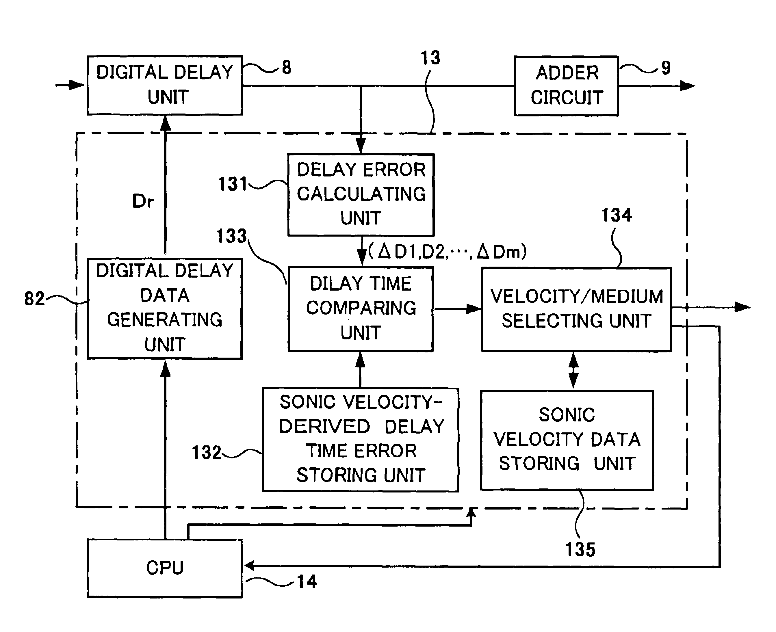
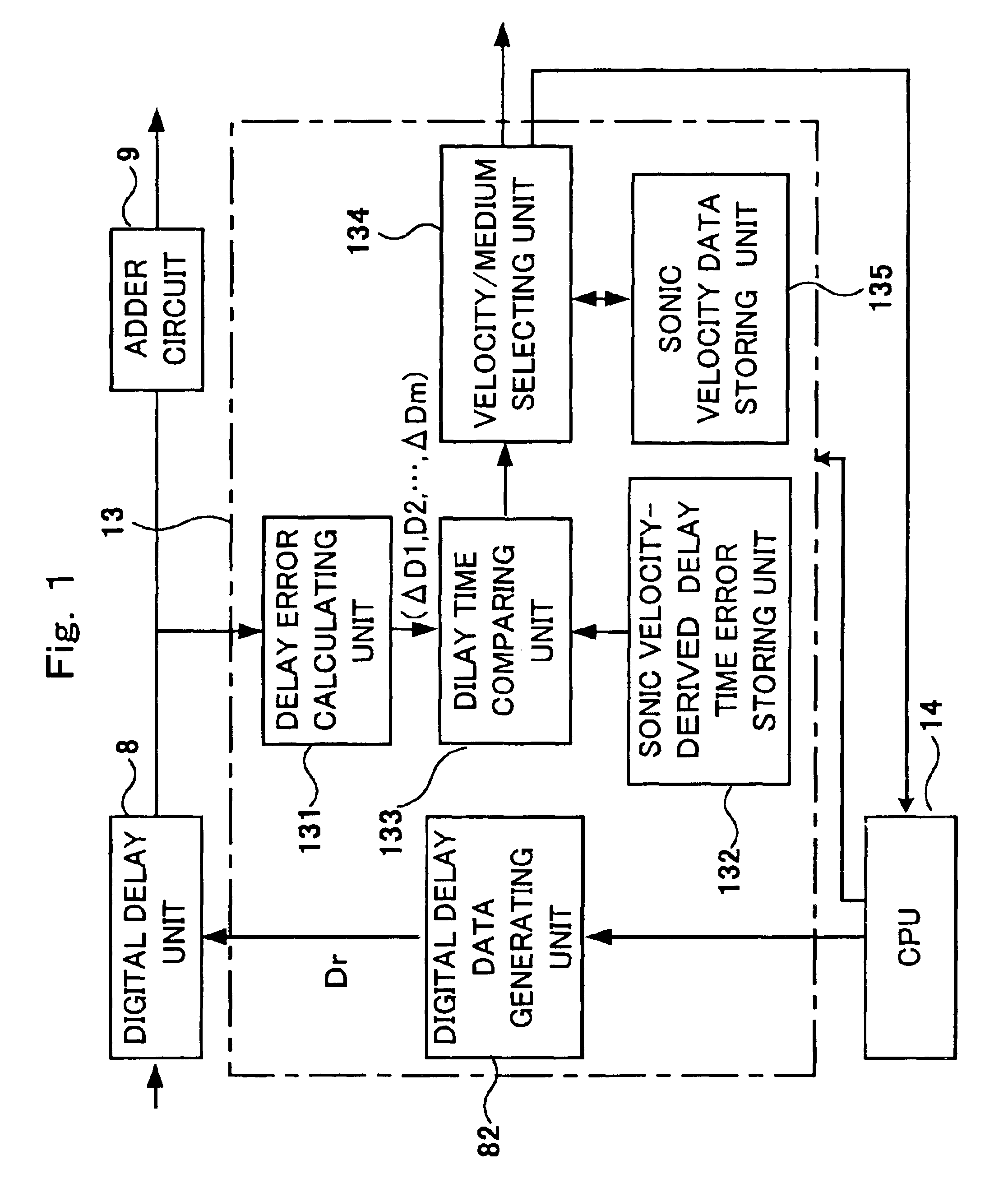
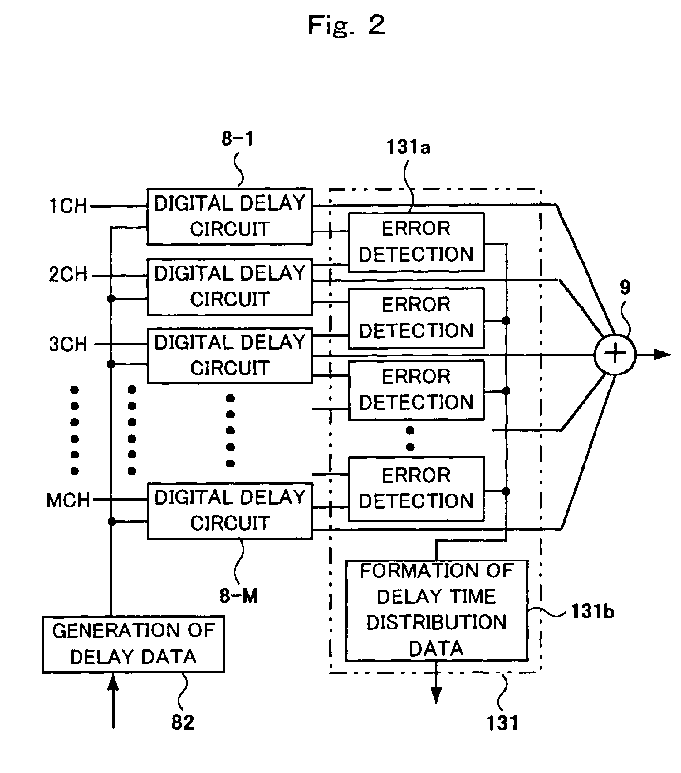
[0038]Hereinafter, an embodiment of the present invention is minutely described using diagrams. FIG. 3 is a block diagram showing the structure of an ultrasonic imaging apparatus. In FIG. 3, 1 is a transmission pulse circuit for generating pulse signals for driving ultrasonic transducers to transmit ultrasound. 2 is a transmission delay circuit for providing each ultrasonic transducer driven by the respective pulse signals output from the transmission pulse circuit 1 with the determined delay time corresponding to each driven transducer. 3 is a transmitting / receiving separation circuit for passing signals from the transmission pulse circuit to the ultrasonic transducer when transmitting ultrasonic waves, and from the ultrasonic transducer side to the receiving circuit side when receiving ultrasonic waves. 4 is a transducer selecting switch circuit for selecting from among the ultrasonic transducers that are arrayed on the ultrasonic probe a transducer group (an aperture) to transmit...


PUM


Abstract

An ultrasonic imaging arrangement performs tentative scanning of ultrasound to the inside of a living body with delay time corresponding to average sonic velocity, calculates delay time error in a delay time error detecting circuit by using received signals from each channel to which delay control has been performed in a digital delay circuit, compares in a delay time comparing unit the calculated data with a plurality of delay time error data corresponding to various sonic velocities using sound velocity as the parameter stored before hand in a sound velocity-derived delay time error storing unit, selects among those sonic velocities the one matching the delay time error data with a sonic velocity selecting unit, and calculates the sonic velocity within the living body. The calculated sonic velocity is fed back to CPU, and the delay data on the delay applied in the ultrasonic scanning is provided to a delay circuit.

Description

FIELD OF THE INVENTION[0001]The present invention relates to an ultrasonic imaging apparatus for examining internal structure of a living body and goods, such as an ultrasonic diagnosis apparatus used for medical diagnosis, and a non-destructive examination apparatus used for non-destructive examination of goods. More particularly, it relates to an ultrasonic imaging apparatus that can obtain images of high quality even when the internal structure of a living body and goods are different in their propagation velocity of ultrasonic wave.BACKGROUND OF THE INVENTION[0002]In a linear scanning type ultrasonic imaging apparatus, ultrasound is received and transmitted by simultaneously driving the elements of an arrayed transducer group forming an aperture, which are selected by a aperture-selecting switch to form the aperture for a plurality of ultrasonic array transducers. Then, by shifting the aperture successively, the inside of the living body or goods is linear-scanned by an ultrason...

Claims


Application Information

Patent Timeline
IPC IPC(8): A61B8/00G01H5/00G01S7/52A61B8/14
CPCA61B8/00G01S7/52049G01S7/52071G01S7/52074
Inventor BABA, HIROTAKASHINOMURA, RYUICHISATO, YUTAKAMIWA, YUICHI
Owner HITACHI MEDICAL CORP
Why Eureka
Try Eureka