Enzyme-mediated modification of fibrin for tissue engineering: incorporation of proteins

a technology of fibrin and enzymes, applied in the field ofmatrices, can solve the problems of altering the release of proteins, and achieve the effects of enhancing neurite outgrowth, promoting angiogenesis, and increasing neurite extension

Inactive Publication Date: 2005-11-01
ETH ZZURICH +1
View PDF14 Cites 25 Cited by
  • Summary
  • Abstract
  • Description
  • Claims
  • Application Information

AI Technical Summary

Benefits of technology

[0005]Synthesis of either of the fusion proteins described above can be accomplished by utilizing molecular biology techniques. To do this, a fusion protein can be created that contains the entire protein sequence of interest with a cross-linking or binding sequence fused onto the amino or carboxyl terminus. This is done at the DNA level, as sequences encoding for either a factor XIIIa cross-linking substrate or a heparin-binding domain can be inserted at the beginning or the end of the codons for the original protein. When these modified proteins are expressed, they will then contain the additional domain of interest at the amino terminus. By using the natural machinery designed for protein synthesis, it becomes possible to synthesize and purify large proteins with high fidelity.Incorporation of Fusion Proteins
[0009]The simplest way to incorporate proteins into fibrin is to attach heparin to the fibrin gels and use the heparin to sequester heparin-binding proteins, such as heparin-binding growth factors. This can be accomplished one of two ways, either by directly coupling a heparin-peptide chimera (where the heparin is chemically attached to a peptide containing a factor XIIIa substrate), or indirectly by cross-linking a heparin-binding peptide into the fibrin gel and binding heparin to this peptide non-covalently (using a bifunctional peptide containing a heparin-binding domain and a factor XIIIa substrate). This heparin can then sequester proteins, such as growth factors with heparin affinity, in the fibrin gel in a manner similar to the way that they are sequestered to the extracellular matrix in nature. Heparin can also protect these factors from proteolytic degradation and prolong their activity until they are released from the matrix.
[0010]Despite their relatively strong affinity for heparin, heparin-binding growth factors dissociate from the matrix on a short time scale. Therefore, a high excess of binding sites is essential to ensure that they do not diffuse far before they bind to the matrix again. This equilibrium also allows for the binding of free growth factor to cell surface receptors that are in close proximity to the site of dissociation. This method of controlled release provides both relatively long-term binding of growth factors and rapid release of growth factors to local cells.
[0012]The attachment of heparin, either covalently or non-covalently to fibrin gels adds a novel functionality to these materials. The attachment of heparin permits the fibrin matrix to bind heparin-binding proteins, including growth factors in a manner which does not harm the protein, and prevents free diffusion of the protein from the gel. This allows for the controlled-release of heparin-binding proteins by one of two mechanisms, either degradation of the gel or binding of the protein to some other high affinity protein, such as a cell surface receptor.
[0030]Heparin (or other polysaccharides such as heparin sulfate or chondroitin sulfate) can be attached to fibrinogen directly using factor XIIIa by constructing a heparin-peptide chimera. This chimera contains two domains, a peptide domain consisting of a factor XIIIa substrate and the polysaccharide domain, such as heparin. These chimeras are made using modified heparin (or another polysaccharide) which contains a unique reactive group at one end to control the site where coupling occurs on the heparin molecule. Through the use of a unique functional group on the peptide, such as a side chain present only on the end of the peptide where coupling is desired, the location of coupling on the peptide can be controlled as well. These chimeras can then be covalently cross-linked to fibrin gels using the sample methods as peptide chimeras, allowing direct attachment of heparin to the fibrin gel.

Problems solved by technology

However, incorporating an enzymatic degradation site alters the release of the protein during proteolysis.

Method used


Image

  • Enzyme-mediated modification of fibrin for tissue engineering: incorporation of proteins
  • Enzyme-mediated modification of fibrin for tissue engineering: incorporation of proteins
  • Enzyme-mediated modification of fibrin for tissue engineering: incorporation of proteins

Examples


example 1

Indirect Coupling of Heparin Via a Heparin-binding Peptide to Attach Growth Factor

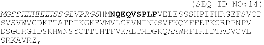
[0036]A peptide chimera containing both a factor XIIIa substrate and a heparin-binding domain is synthesized by standard solid phase synthesis. A sample peptide is one containing the following sequence, dLNQEQVSPK(βA)FAKLAARLYRKA (SEQ ID NO:12), where the N-terminus of the peptide contains the factor XIIIa substrate and the sequence in italics contains a modified peptide from the heparin-binding domain of ATIII (dL denotes dansyl leucine, which is used to allow detection of the peptide by fluorescence).

[0037]Size exclusion chromatography was used to determine the amount of peptide cross-linked to fibrin gels using the previously developed incorporation method. A bi-domain peptide containing the heparin-binding domain from antithrombin III and a fluorescent label was incorporated into fibrin gels during polymerization. The free peptide was washed from the gels, and the fibrin network was degraded with p...

example 2

Synthesis of Heparin-peptide Chimeras

[0038]A heparin-peptide chimera is synthesized by coupling a peptide, containing the factor XIIIa substrate on the N-terminus and a poly-lysine on the C-terminus, to a heparin oligosaccharide, with a unique aldehyde group on one end, via reductive amination. A peptide with the following sequence, dLNQEQVSPLKKKG (SEQ ID NO:13), is synthesized by standard solid phase peptide chemistry. The heparin oligosaccharides are made by standard nitrous acid degradation of heparin, resulting in the formation of an aldehyde on the reducing terminal of the cleaved oligosaccharide. During coupling, the ε-amino group of the lysine side chain attacks the aldehyde on the reducing end of the heparin oligosaccharide to form a Schiff base. The Schiff base is then reduced to form a stable product. A sample coupling protocol is given below.

Coupling Protocol:

[0039]1) Dissolve 1.8 mM of peptide and 1.8 mM of nitrous acid degraded heparin in 50 mM borate buffer, pH 9. Reac...

example 3

Degradable Sites in Fusion Protein and in Peptide Chimera

[0061]Fusion proteins or peptide chimeras, which are cross-linked to fibrin gels, may be further modified to contain a degradable site between the attachment site (i.e., factor XIIIa substrate or heparin-binding domain) and the bioactive protein (i.e., growth factor or enzyme). These sites may be degradable either by non-specific hydrolysis (i.e., an ester bond) or they may be substrates for specific enzymatic (either proteolytic or polysaccharide degrading) degradation. These degradable sites allow the engineering of more specific release of bioactive factor from fibrin gels. For example, degradation based on enzymatic activity allows for the release of bioactive factors to be controlled by a cellular process rather than by diffusion of the factor through the gel.

[0062]The degradation sites allow the bioactive factor to be released with little or no modification to the primary protein sequence, which may result in higher acti...


PUM

PropertyMeasurementUnit
bioactiveaaaaaaaaaa
affinityaaaaaaaaaa
non-covalent interactionsaaaaaaaaaa

Abstract

Disclosed are materials that may be used in the design of improved devices and wound treatment platforms though covalent and / or non-covalent attachment of bioactive proteins. The proteins comprise any variety of cell growth and / or healing promoting proteins, such as growth factor. The incorporation of these whole proteins may be designed to provide controlled release thereof in a biological system through further use of enzyme degradation sites. Heparin-binding protein or fusion proteins synthesized to contain a heparin-binding domain are two mechanisms that may be used in providing these properties to a matrix, such as a fibrinogen matrix. The proteins will be used to provide enhanced healing in various tissues including vasculature, skin, nerve, and liver. The materials disclosed will be used to enhance would?? Healing and other generative processes by engineering the fibrin gel to contain appropriate proteins with specifically designed release and / or degradation characteristics.

Description

[0001]This application is a continuation of U.S. application Ser. No. 09 / 141,153 filed Aug. 27, 1998, now abandoned.FIELD OF THE INVENTION[0002]The present invention relates generally to the fields of matrices that are modified to enclose particularly designed fusion proteins. More particularly, it concerns the use of fusion proteins that include internal degradation sites and / or enzymatic cleavage sites. Artificial matrices may be designed having desired degradation rates, as well as to include particular active biological molecules, such as growth factors or enzymes.BACKGROUND OF THE INVENTION[0003]It has been demonstrated that bi-domain peptides, which contain a factor XIIIa substrate sequence and a bioactive peptide sequence, can be cross-linked into fibrin gels and furthermore, that this bioactive peptide retains its cellular activity in vitro. While peptides can partially mimic the bioactivity of the whole protein from which they are derived, this bioactivity is usually lower ...

Claims


Application Information

Patent Timeline
Patent Type & Authority Patents(United States)
IPC IPC(8): C07K14/435C07K14/475
CPCC07K14/475
Inventor HUBBELL, JEFFREY A.SCHENSE, JASON C.SAKIYAMA, SHELLY E.
Owner ETH ZZURICH
Why Eureka
Try Eureka