
System for use in displaying images of a body part

a body part and image technology, applied in the field of system for displaying images of body parts, can solve the problems of large devices, high cost, and encircling the body part being imaged, and achieve the effect of convenient us

Inactive Publication Date: 2005-12-20
SAINT LOUIS UNIVERSITY +1
View PDF211 Cites 231 Cited by
  • Summary
  • Abstract
  • Description
  • Claims
  • Application Information

AI Technical Summary

Benefits of technology

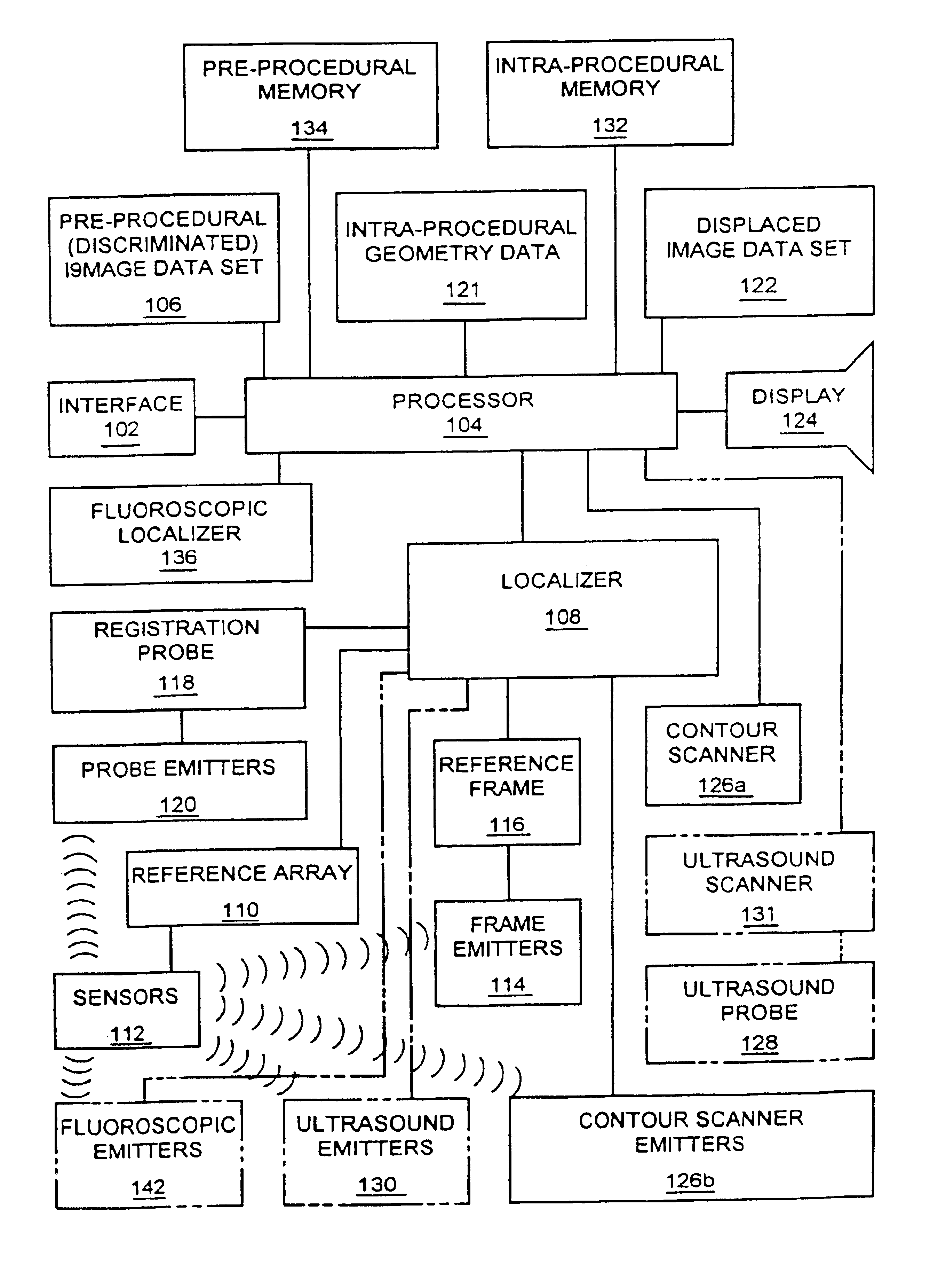
[0009]It is a further object of this invention to provide a system which can localize multiple rigid bodies that move with respect to each other between imaging and a procedure and provide a display during the procedure of the bodies in their displaced positions.
[0013]It is a further object of this invention to provide a system for use during image guided medical and surgical procedures which is easily employed by the doctor or surgeon conducting the procedure.

Problems solved by technology

The majority of the advances in imaging involve devices which tend to be large, encircle the body part being imaged, and are expensive.
Although the images produced by these devices depict the body part under investigation with high resolution and good spatial fidelity, their cost usually precludes the dedication of a unit to the performance of procedures.

Method used


Image

  • System for use in displaying images of a body part
  • System for use in displaying images of a body part
  • System for use in displaying images of a body part

Examples


Embodiment Construction

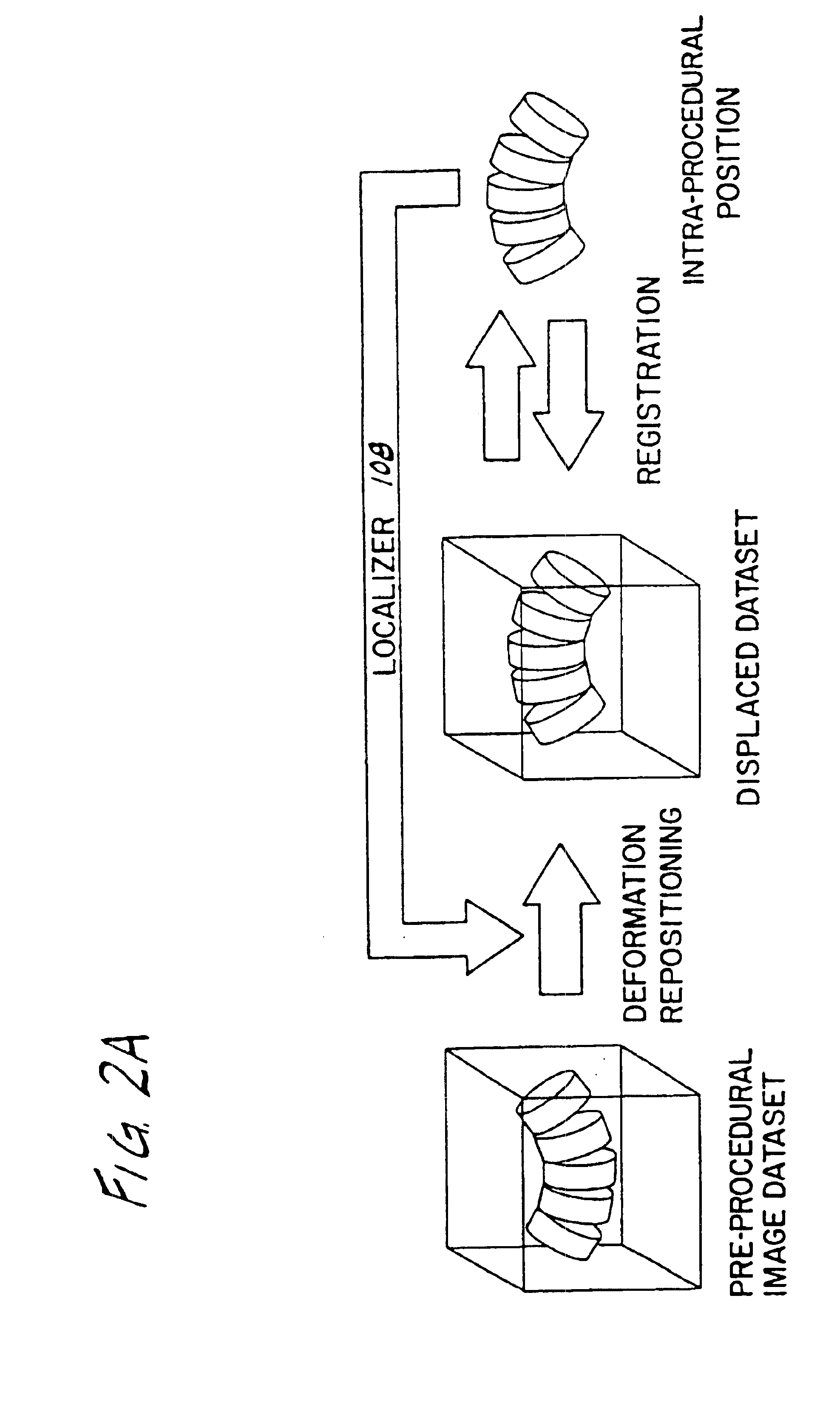
[0037]Referring to FIG. 2A, an overview of operation of one preferred embodiment of the system according to the invention is illustrated. Prior to a particular procedure, the body elements which will be part of the procedure are scanned to determine their alignment. For example, the alignment may be such as illustrated in FIG. 3 wherein body elements 10, 20, and 30 are more or less aligned in parallel. These body elements may be bones or other rigid bodies. In FIG. 3, three-dimensional skeletal elements 10, 20, 30 are depicted in two dimensions as highly stylized vertebral bodies, with square vertebra 11, 21, 31, small rectangular pedicles 12, 22, 32, and triangular spinous processes 13, 23, 33. During imaging, scans are taken at intervals through the body parts 10, 20, 30 as represented in FIG. 3 by nine straight lines generally referred to be reference character 40. At least one scan must be obtained through each of the body elements and the scans taken together constitute a three...


PUM


Abstract

A system for use during a medical or surgical procedure on a body. The system generates a display representing the position of two or more body elements during the procedure based on a reference image data set generated by a scanner. The system produces a reference image of a body elements, discriminates the body elements in the images and creates an image data set representing the images of the body elements. The system produces a density image of the body element. The system modifies the image data set according to the density image of the body element during the procedure, generates a displaced image data set representing the position and geometry of the body element during the procedure, and compares the density image of the body element during the procedure to the reference image of the body element. The system also includes a display utilizing the displaced image data set generated by the processor to illustrate the position and geometry of the body element during the procedure. Methods relating to the system are also disclosed.

Description

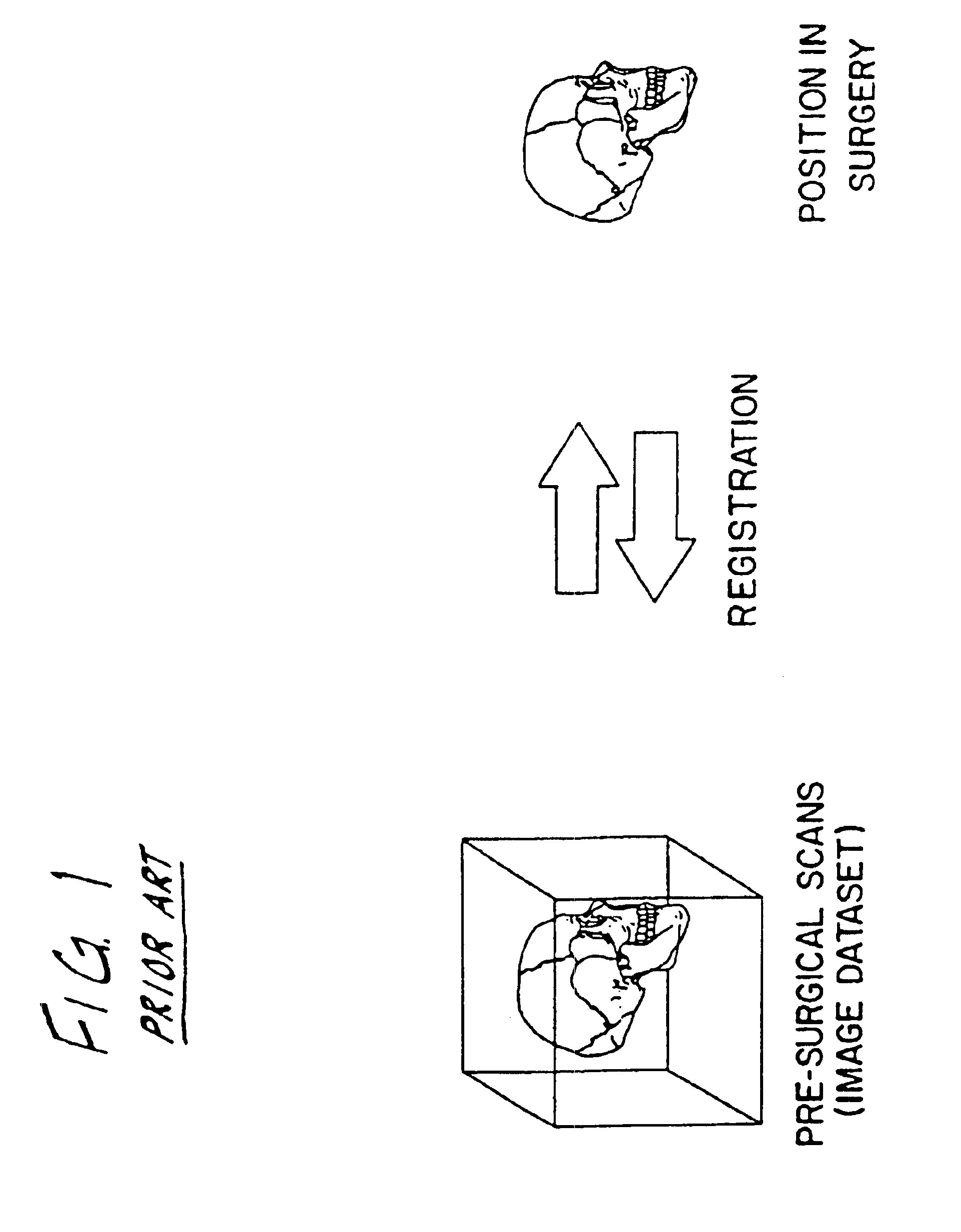
[0001]This application is a continuation of U.S. patent application Ser. No. 09 / 398,313, filed on Sep. 20, 1999, now U.S. Pat. No. 6,434,415, which is a continuation of U.S. patent application Ser. No. 08 / 931,654 filed on Sep. 16, 1997, now U.S. Pat. No. 6,347,240, which is a continuation of U.S. patent application Ser. No. 08 / 319,615, filed on Oct. 7, 1994, now abandoned.BACKGROUND OF THE INVENTION[0002]The invention relates generally to systems which generate images during medical and surgical procedures, and in particular, a system for generating images during medical and surgical procedures based on a scan taken prior to the procedure.[0003]Image guided medical and surgical procedures comprise a technology by which images, obtained either pre-procedurally or intra-procedurally (i.e., prior to or during a medical or surgical procedure), are used to guide a doctor during the procedure. The recent increase in interest in this field is a direct result of the recent advances in imagi...

Claims


Application Information

Patent Timeline
IPC IPC(8): A61B5/00A61B5/055A61B5/06A61B5/107A61B6/00A61B6/12A61B8/00A61B17/00A61B17/88A61B19/00
CPCA61B5/0064A61B5/0073A61B5/06A61B5/1077A61B6/12A61B6/501A61B8/00A61B8/5238A61B19/20A61B19/52A61B19/5244A61B5/055A61B19/203A61B19/50A61B19/54A61B2017/00022A61B2019/5238A61B2019/5251A61B2019/5268A61B2019/5272A61B2019/5276A61B2019/5287A61B2019/5289A61B2019/5425A61B2019/5429A61B2019/5445A61B2019/5454A61B2019/5483A61B5/061A61B5/062A61B34/10A61B34/20A61B90/10A61B90/14A61B90/36A61B90/39A61B2034/2051A61B2034/2068A61B2034/2072A61B2090/363A61B2090/364A61B2090/376A61B2090/378A61B2090/3925A61B2090/3929A61B2090/3945A61B2090/3954A61B2090/3983
Inventor FOLEY, KEVIN T.BUCHOLZ, RICHARD D.SMITH, KURT
Owner SAINT LOUIS UNIVERSITY
Learn More